
Introduction
Foot and ankle care · Arch problems
Flat Feet in Adults: Assessment and Treatment Options

Dr Sean LeoOrthopaedic Surgeon, Singapore
Clinic enquiries · Not an emergency service
Illustrative image

Flat feet are common; a newly flattening arch in an adult deserves understanding.
Flat feet are common; a newly flattening arch in an adult deserves understanding.
Understand the problem
Flat feet are common; a newly flattening arch in an adult deserves understanding.
Flat feet (adult-acquired flatfoot) are common and often painless. An arch that newly flattens in adult life can signal a problem with the tendon that holds the arch. In the early stages, most people are managed without surgery. An assessment separates a harmless shape from a changing one.[1][5]
This page covers flat feet (adult-acquired flatfoot) in adults. Flat feet in children are a different discussion and are not covered here.
Seeing a specialist about a foot or ankle problem does not mean surgery has already been decided. An assessment helps you weigh reasonable options.
OrthoKinetics is Dr Sean Leo’s professional brand and patient-education platform. Clinical consultations and treatment are provided through OrthoSports Orthopaedic & Sports Surgery. Dr Sean Leo is a specialist orthopaedic surgeon who cares for foot and ankle problems.
What’s covered on this webpage
Follow the route in order, or jump to the stop you need. Stops you have read are marked.
The arch is held by a tendon and ligaments working together.
The arch is held by a tendon and ligaments working together.
The arch is held by a tendon and ligaments working together.
The arch is the gap under the inside of your foot. Bone gives it a frame. Soft tissue holds it up.[1][5]
The main support is the posterior tibial tendon (the tendon behind the inner ankle that holds up the arch). Ligaments under the arch help, including the spring ligament, a major stabiliser of the arch while you walk.[2][5]
Soft tissue can change gradually if it struggles. That is why the adult story usually starts with the tendon.

In adults, a flattening arch usually begins with the tendon that holds it.
In adults, a flattening arch usually begins with the tendon that holds it.
In adults, a flattening arch usually begins with the tendon that holds it.
Many flat feet are lifelong and cause no trouble.[1][5]
The adult-acquired pattern is different. It is most commonly believed to begin when the posterior tibial tendon is inflamed or torn and may not support the arch. The ligaments under the arch can then give way too.[2][5]
The result is a three-dimensional change, not just a lower arch. The heel drifts outward, the front of the foot turns outward and the arch lowers.[1]
Two other names are posterior tibial tendon dysfunction (PTTD), the older name, and progressive collapsing foot deformity (PCFD), chosen by an international expert group in 2020.[1] The new name did not change the treatment evidence.
Several general-health factors are linked with it:[3]
- age-related wear;
- inflammatory arthritis;
- high blood pressure;
- diabetes;
- obesity.
Less often, a single injury tears the tendon.[3] These are associations, not blame. A mild early change often stays stable. It can progress, but not always.[1][5]
The early signs are a tired inner ankle and a slowly changing shape.
The early signs are a tired inner ankle and a slowly changing shape.
The early signs are a tired inner ankle and a slowly changing shape.
The changes are often slow and easy to ignore.[2][3]
| What you might notice | What it can mean |
|---|---|
| Ache or swelling along the inner ankle or arch | The arch-supporting tendon may be struggling |
| The arch looks lower than it used to | The supporting structures may be gradually failing |
| The heel drifts outward; from behind, more toes show on the outer side | The heel bone is tilting, part of the three-dimensional change |
| Rising onto one foot has become harder | The tendon may not be holding the arch as it should; a clue, not a diagnosis |
Two signs have names. The “too many toes” sign is what someone sees standing behind you: more toes show on the outer side of each foot, because the heel has drifted outward. The single heel raise asks you to rise onto the ball of one foot. Difficulty can suggest the tendon is struggling. It is a clue, not a diagnosis.[2]

Can flat feet affect my knees or hips
Can flat feet affect my knees or hips
Can flat feet affect my knees or hips?
Flat feet have been linked with knee pain in a study of older adults. This shows a link, not proof that one causes the other.[8]
Care can include suitable footwear or custom orthotics, foot and ankle exercises, and in certain cases surgery.[3][5][7]
Watch: Your foot arch might be the reason for your knee pain
Flexible and rigid flat feet are managed differently.
Flexible and rigid flat feet are managed differently.
Flexible and rigid flat feet are managed differently.
Flat feet are not all the same. The most useful split is flexible versus rigid.[1][5]
| Flexible | Rigid | |
|---|---|---|
| What you see | The arch returns when you sit or lift the foot, and lowers when you stand | The shape stays flat however the foot is held |
| What usually follows | Supports, footwear and exercise are often tried first | Supports may still ease symptoms; surgery, if needed, is a different operation |
The range is wide. It runs from a subtle change with mild symptoms to a painful foot that works poorly.[5]

When should flat feet be assessed?
When should flat feet be assessed?
When should flat feet be assessed?
Arrange an assessment if:
- your arch has newly flattened in adult life;
- inner ankle ache or swelling persists or keeps returning;
- your heel has drifted outward, or the “too many toes” look is new;
- rising onto one foot has become noticeably harder;
- the change limits walking, work or recreation.
Safety note: This webpage is not an emergency service. The signs that need urgent help are listed at the end of this page. No list on this page can rule out a serious problem remotely.
Assessment looks at the whole foot, not one test.
Assessment looks at the whole foot, not one test.
Assessment looks at the whole foot, not one test.
Assessment starts with your story: when the shape changed, what hurts and what you can no longer do.
The examination looks at the arch and heel while you stand, the view from behind, and how you walk. The single heel raise is a clue, not a verdict.[2]
Are scans needed?
Scans are added only when they answer a useful question. X-rays, usually taken standing, and sometimes an MRI scan, can add detail. A scan supports the examination and does not choose treatment by itself.

Support comes first, and it helps many people.
Support comes first, and it helps many people.
Support comes first, and it helps many people.
In the early stages, most people are treated without surgery, using orthotics and braces.[5] Support usually combines:
- Footwear. Sensible, supportive footwear is the base of foot care.[7] See what to look for in a shoe.
- Insoles (orthoses). They support the arch. The video below explains the types.[2][5]
- Braces. An ankle-foot orthosis is a brace that extends above the ankle, used for more advanced stages. The evidence is supportive but limited.[2]
- Exercise. A structured programme of stretching and progressive strengthening.[3]
In one trial of 36 adults with early disease, a 12-week programme of custom orthoses, stretching and resistive exercise improved pain and function in every group. The eccentric strengthening group (strengthening while the muscle lengthens) improved most.[3] The trial did not measure whether the arch changed shape or flattening stopped.
A first pair of insoles that does not help does not mean surgery is next.
Watch: Orthotics: insoles and arch supports explained
What does the research say about bracing?
In one group of 64 adults followed for an average of 27 months, 56 (87.5%) were successfully managed without surgery using bracing, physiotherapy and anti-inflammatory medicine. Bracing was linked with success. A tendon split-tear on MRI was linked with non-surgical care not working.[4] This was a review of records with no comparison group, and the group had a high rate of obesity. The figure describes that group, not you.
When is surgery part of the conversation
When is surgery part of the conversation
When is surgery part of the conversation?
Surgery is discussed when orthotics and braces do not provide relief.[5] The aim is to help pain and function, not to give you a new foot. It is usually very complex, and most people still notice some limitation in activity afterwards.[5]
Structure / Patient / Goal
Structure / Patient / Goal
Deciding together
Structure / Patient / Goal
S
Structure: what is changing in the foot?
Structure is which tissues are struggling and whether the foot is flexible or rigid.
P
Patient: how does it affect you?
Patient is you as a whole person: pain, activity, general health and preferences.
G
Goal: what are you hoping to do?
Goal is what you want to return to, such as comfortable walking, work, recreation or sport.
Your plan
No single factor decides the plan.
Support, realignment and fusion answer different problems.
Support, realignment and fusion answer different problems.
Support, realignment and fusion answer different problems.
If an operation is needed, it follows the deformity.[5][6]
| Deformity | Operation family |
|---|---|
| Early disease | Tendon clean-up or repair |
| Flexible deformity | Bone realignment with tendon transfer |
| Rigid deformity | Fusion of hindfoot joints |
The more flexible the foot, the more an operation can preserve. The stiffer it is, the more it must stabilise.[5][6]
Examination, imaging and Structure / Patient / Goal guide the choice, made with you at assessment.
What flat foot surgery involves, in outline.
What flat foot surgery involves, in outline.
What flat foot surgery involves, in outline.
Tendon surgery. In early disease, the tendon is cleaned up or repaired.[5]
Realignment. For a flexible deformity, the heel bone can be moved inward (a calcaneal osteotomy) and a neighbouring tendon transferred to take over. Sometimes the outer side of the foot is lengthened too.[5][6]
Fusion. For a rigid deformity, stiff hindfoot joints are joined in a corrected position so they stop hurting. Double or triple fusion is frequently needed.[6]
Operations may be combined, and the detail is individual. Most people still notice some limitation in activity afterwards.[5]
Recovery is measured by milestones, not dates.
Recovery is measured by milestones, not dates.
Recovery
Recovery is measured by milestones, not dates.
With non-surgical care, a structured programme runs in weeks to months. The trial programme above lasted 12 weeks. That describes the trial, not your timetable.[3] Progress is judged by symptoms and function.
After surgery, recovery depends on the operation and the person.

Risks and honest limits.
Risks and honest limits.
Risks and honest limits.
Each path has limits.
- Progression. A flat foot can stay stable for years. It can also progress, so review matters more than worry.[1][5]
- Support. The trial evidence measured pain and function, not arch correction or halting progression.[3]
- Cohort finding. In one group, a tendon split-tear on MRI was linked with bracing not working. It is not a personal prediction.[4]
- Surgery. Most people still notice some limitation in activity afterwards.[5]

OrthoKinetics Recovery Reflection
Recovery reflection
If a change happens slowly and barely hurts, what makes noticing it worth acting on?
You have lived in your feet longer than anyone. A slow change you noticed is real information.
What would it mean to say what you have noticed out loud?
It means bringing your own long observation into the room. Attention is care, neither alarm nor neglect.
Illustrative image

 With you through the seasons
Meet Dr Sean Leo
Meet Dr Sean Leo
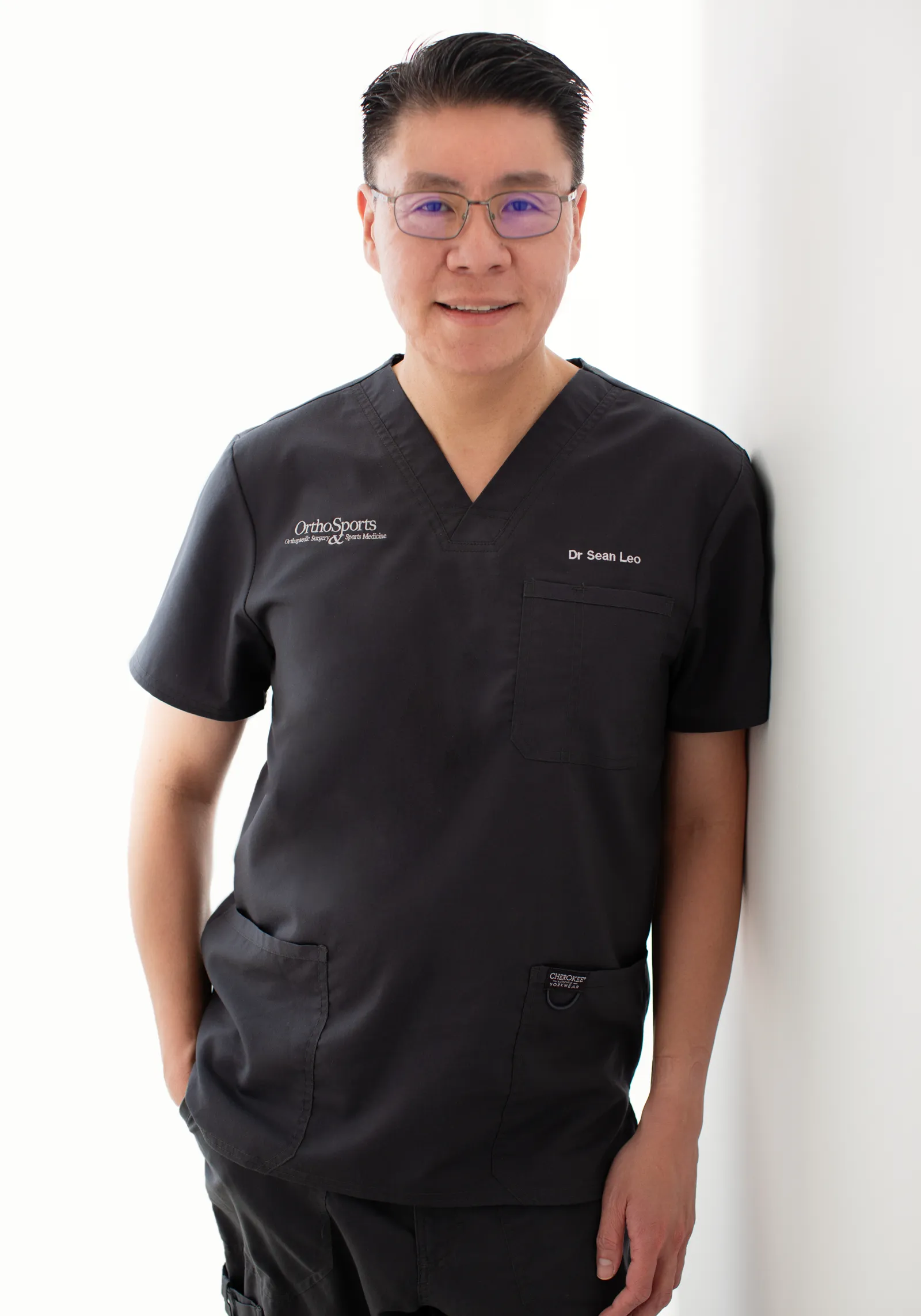
A consultant orthopaedic surgeon whose clinical focus is the knee, sports injuries and the lower limb.
- Dr Sean Leo is a consultant orthopaedic surgeon. OrthoKinetics is his professional brand; clinical services are provided through OrthoSports Orthopaedic & Sports Surgery at Mount Elizabeth Novena Specialist Centre and Connexion, Farrer Park Hospital.
- His clinical interests include sports injuries, including those in adolescent athletes; ligament reconstruction; realignment surgery; cartilage repair and joint-preservation approaches; knee replacement; and technology-assisted hip and knee surgery.
- The consistent aim is to return people to meaningful function and activity while avoiding unnecessary downtime.
- Qualifications: MBBS, MRCS (Ed), MMed (Ortho), FRCSEd (Ortho).
More detail: Meet Dr Sean Leo
- Training and career: Dr Leo trained in medicine at the National University of Singapore and served in the Singapore Armed Forces Medical Corps from 2000 to 2012, where his work included the care of soldiers in physically demanding roles. He completed postgraduate orthopaedic training in 2009 and a one-year clinical fellowship at Royal North Shore Hospital in Sydney, Australia, before joining Changi General Hospital as an orthopaedic consultant in 2012, where he later held the positions of Senior Consultant and Director of the Hip and Knee Service. With seniority, his educational contribution has evolved towards mentorship and support for junior doctors.
Questions people often ask
Common flat feet questions
Common flat feet questions
Why are my feet flat now when they were not before?
An adult-acquired change is most commonly believed to begin when the posterior tibial tendon stops supporting the arch. The ligaments under the arch can then give way. It is not anyone’s fault.[2][5]
Can flat feet cause knee or hip pain?
Flat feet have been linked with knee pain in a study of older adults. This shows a link, not proof that one causes the other.[8] That research looked at knee pain; it did not test the hip. Many other things also cause knee and hip pain.
Do I need custom insoles?
Many people benefit from insoles that support the arch. Whether yours should be custom-made is decided at assessment.[2][5]
Can exercise fix flat feet?
Structured stretching and strengthening improved pain and function in a 12-week trial. That trial did not measure whether the arch changed shape.[3]
Is flat foot painful, or is it just a shape?
Often it is only a shape and causes no trouble. A new change, pain or limits on what you do are the reasons to be assessed.[5]
When should flat feet be checked?
When the arch newly flattens, inner ankle ache or swelling persists, the heel drifts outward, or the change limits you. Stable, lifelong, painless flat feet usually need no check.
What is the tendon behind the inner ankle, and what does it do?
It is the posterior tibial tendon. It runs behind the inner ankle and supports the arch. When it struggles, the arch can lower.[2][5]
Can flat feet get worse over time?
They can, but not always. A mild early change often stays stable for years. Review is the proportionate response, not worry.[1][5]
What is the single heel raise test?
You rise onto the ball of one foot while a clinician watches. Difficulty can suggest the arch-supporting tendon is struggling. It is a clue, not a diagnosis.[2]
Costs and insurance
Costs and insurance
Practicalities
Cost, MediSave and insurance

Ask the clinic team for current information about fees, panel status, pre-authorisation and expected payment arrangements. Insurance coverage and out-of-pocket costs depend on the individual policy and the insurer’s decision; no coverage is guaranteed.
A cost estimate for surgery can be given only after a consultation and assessment, once a surgical plan has been developed. Each individual condition is unique — implants used, time taken, and other factors contribute to the overall estimate. The estimate will be provided to you by the clinic should you decide on surgical management, and will also be used to assist in obtaining pre-authorisation from insurance where applicable.
A cost estimate for surgery can be given only after a consultation and assessment, once a surgical plan has been developed. A surgical estimate separates surgeon, anaesthetist, hospital or facility, implant and consumable costs, and the MOH publishes national fee benchmarks for common procedures. For how costs, MediSave and insurance work in Singapore, see Costs, insurance and MediSave, explained.
Insurance panels. Dr Sean Leo, Orthopaedic Surgeon, is on the specialist panels of Great Eastern (GE Panel), HSBC Life (HSBC Life Shield) and Singlife (Singlife Shield), and on the Prudential Panel. AXA Insurance Singapore became part of HSBC Life in 2023, and former AXA Shield plans are now HSBC Life Shield plans.
Corporate health plans. Dr Leo is on the panels of both Alliance Medinet and iXchange, which administer medical benefits for many company health plans. If your company plan uses either panel, you can see Dr Leo as a panel doctor.
If your company uses a different panel. Some company plans still let you see a doctor of your choice outside their panel, then claim reimbursement. This varies from plan to plan, as may the co-payment, so please check your benefits with your company’s HR department.
Panel arrangements change from time to time, so please confirm with your insurer or your company’s plan administrator before your appointment.
Dr Leo is on the panels shown. If your insurer is not listed, you may ask whether co-management with an Orthosports surgeon partner on that panel is clinically and administratively appropriate. Orthosports surgeon partners are collectively on the panels of all Shield plans.
Specific benefits, pre-authorisation, reimbursement and cashless treatment depend on the individual policy and insurer approval and are not guaranteed.
Ready to book? The appointment section is at the end of this page.
Arrange a consultation: the appointment section is further down this page.
Sources
References
References
- Classification and Nomenclature: Progressive Collapsing Foot Deformity. Foot Ankle Int. 2020. PMID 32856474.
- American College of Foot and Ankle Surgeons. Clinical Consensus Statement: Appropriate Clinical Management of Adult-Acquired Flatfoot Deformity. J Foot Ankle Surg. 2020;59:347-355. Available at: https://www.acfas.org/ACFAS/media/ACFAS_Media/Adult-Acquired-Flatfoot-Deformity-CSS.pdf
- Kulig K, et al. Nonsurgical management of posterior tibial tendon dysfunction with orthoses and resistive exercise: a randomized controlled trial. Phys Ther. 2009;89(1):26-37. PMID 19022863. doi:10.2522/ptj.20070242.
- Nielsen MD, et al. Nonoperative care for the treatment of adult-acquired flatfoot deformity. J Foot Ankle Surg. 2011;50(3):311-314.
- American Academy of Orthopaedic Surgeons (AAOS). Progressive Collapsing Foot Deformity (Flatfoot). OrthoInfo. Available at: https://orthoinfo.aaos.org/en/diseases–conditions/posterior-tibial-tendon-dysfunction/
- Progressive Collapsing Foot Deformity – Flatfoot. Springer textbook chapter. Available at: https://link.springer.com/article/10.1007/978-3-030-95738-4_24
- Health Promotion Board, Singapore. Podiatry. HealthHub. Available at: https://www.healthhub.sg/support-and-tools/support-and-care-programmes/podiatry
- Gross KD, Felson DT, Niu J, Hunter DJ, Guermazi A, Roemer FW, Dufour AB, Gensure RH, Hannan MT. Association of flat feet with knee pain and cartilage damage in older adults. Arthritis Care Res (Hoboken). 2011;63(7):937-944. doi:10.1002/acr.20431. PMC3087845. Available at: https://pmc.ncbi.nlm.nih.gov/articles/PMC3087845/
Final next step
Final next step
Next step
Final next step
Reading about a problem is a starting point. An assessment brings together the history, an examination where needed and any useful tests, and helps you and your clinician decide what to do next. How much physical examination is needed is a matter of clinical judgement. It may be limited when pain makes an examination unreliable, or when imaging clearly shows a problem that matches the symptoms, such as a fracture seen on an X-ray or an MRI scan that clearly shows an injury consistent with the symptoms. The Singapore Medical Council and the UK General Medical Council both allow remote medical consultations, where a physical examination is not possible, with safeguards: the doctor must be clear about the limits of an opinion formed without an examination, and whether an in-person examination is needed, or referral to other services, is left to the clinical judgement of the attending doctor.
Signs that need urgent help
Some symptoms should not wait for a routine appointment. Each item below says where to go.
After an injury
- The ankle or foot sits at an odd angle, a bone is showing, there is a deep cut, you are in severe pain, or the toes look blue or white or feel numb. Go to an emergency department or call 995.
- You felt a sudden pop or sharp pain at the back of the heel or calf, and pushing off or rising onto your toes is difficult. Arrange an urgent same-day assessment.
- It is very painful or the pain is getting worse, the swelling or bruising is large or spreading, or it hurts to put weight on the ankle or foot. Arrange an urgent same-day assessment.
At any time
- A joint, such as the ankle, is suddenly very painful, hot, red and swollen, especially with a fever or feeling generally unwell. Go to an emergency department or call 995.
- One calf or thigh is newly painful and swollen. Arrange an urgent same-day assessment. If you also feel short of breath or have chest pain, call 995.
If you have diabetes
- You have a foot sore and a fever or feel very unwell, or the foot is cold, pale or blue. Or skin on the foot or toes has turned black, or a sore looks deep, or there is pus or redness spreading up the foot. Go to an emergency department or call 995.
- You have a new foot sore, or a foot that is hot, swollen, changed in colour or leaking fluid, without the signs above. Arrange an urgent assessment within 24 hours.
If you are admitted by the doctor at the urgent care centre of one of these private hospitals, the Parkway Pantai hospitals (Mount Elizabeth Orchard, Mount Elizabeth Novena, Gleneagles and Parkway East), Farrer Park Hospital and Thomson Medical Centre, you may ask for Dr Sean Leo to be your attending orthopaedic surgeon. If you are instead discharged and referred for orthopaedic follow-up, you may contact the clinic to arrange an appointment. Both are subject to the hospital’s admission processes and to Dr Leo’s availability.
Was this page helpful?
Arrange a consultation
Appointments are arranged through the OrthoSports clinic team. Send your details below, or message or call the clinic.
Leave your details and the clinic team will reply to arrange a time.
Message the clinic on WhatsApp
-
38 Irrawaddy Road, #10-41 Mount Elizabeth Novena Specialist Centre, Singapore 329563 Map for Orthosports@Novena (opens Google Maps)
-
1 Farrer Park Station Road, #15-17 Connexion, Singapore 217562 Map for Orthosports@Connexion (opens Google Maps)
WhatsApp is for clinic enquiries only. It is not an emergency service and is not monitored for urgent care. Website or messaging contact is not an emergency-response service. For a medical emergency, use your local emergency service.
- OrthoKinetics does not provide emergency or urgent care through this webpage or its WhatsApp contact.