
Introduction
Knee care · Kneecap instability
Kneecap realignment surgery (tibial tubercle osteotomy): when a bony correction restores patellar stability

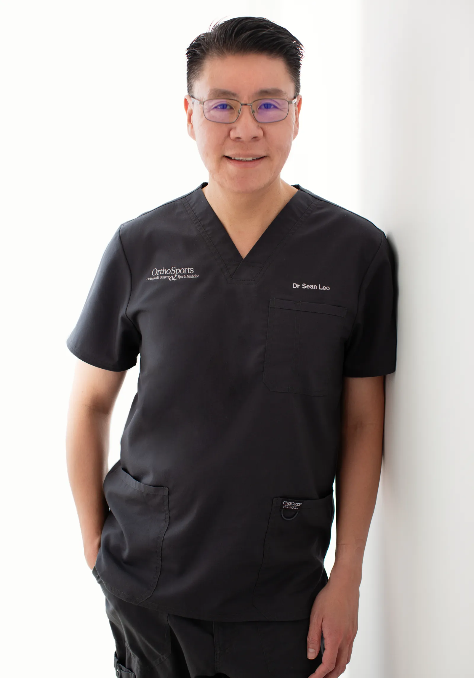
Dr Sean LeoOrthopaedic Surgeon, Singapore
Clinic enquiries · Not an emergency service
Illustrative image
Kneecap realignment in brief
Kneecap realignment in brief
Kneecap realignment in brief
Kneecap realignment — clinically, a tibial tubercle osteotomy (TTO) — is a bony correction for patellar instability. It is considered when the kneecap’s tracking is driven by bone and alignment issues rather than by the ligament alone. Surgery is not automatic: a soft-tissue option (MPFL reconstruction) also exists, and assessment decides which fits, and whether both are combined.
What’s covered on this webpage
Follow the route in order, or jump to the stop you need. Stops you have read are marked.

Understanding patellar instability
Understanding patellar instability
Understand the problem
Understanding patellar instability
Patellar instability is when the kneecap slips partly (subluxation) or completely (dislocation) out of its groove. It usually reflects a combination of anatomy — a shallow groove, a high-riding kneecap, an abnormal TT-TG distance (how far the kneecap tendon’s attachment on the shin bone sits from the groove), or lax ligaments — and an injury that tears the MPFL. Two operations treat it: soft-tissue reconstruction (MPFL) or bony realignment (tibial tubercle osteotomy).
The kneecap (patella) is a small bone that glides in a groove at the end of the thigh bone. A band of tissue called the medial patellofemoral ligament, or MPFL, is the main soft-tissue restraint that stops the kneecap drifting outward. In the early part of bending, it contributes roughly 50–70% of the total inward restraining force — one classic biomechanical study measured about 60% at 20 degrees of flexion. The groove itself and the inner thigh muscle (the VMO) also help hold the kneecap in place.
Dislocation, subluxation and chronic instability
A dislocation is when the kneecap comes fully out of its groove. A subluxation is a partial slip — the kneecap moves out and back without coming all the way out. Chronic instability is the ongoing sense that the kneecap may give way again. When the kneecap dislocates laterally, it tears or stretches the MPFL in more than 9 in 10 cases. The event can also bruise or chip the joint cartilage.
Why the kneecap keeps giving way (the “train on tracks” groove)
Think of the kneecap as a train and the groove as its track. A deep, well-shaped track keeps the train centred. If the track is shallow, or the kneecap rides too high to sit in it, or the attachment point below pulls sideways, the train slips more easily — especially after the ligament that normally holds it in has been stretched or torn. That is why one dislocation can leave a knee that keeps feeling as if it will give way.
What makes a kneecap unstable
What makes a kneecap unstable
What makes a kneecap unstable? (structural contributors)
Instability is usually the result of anatomy and injury together, not a single cause. These factors often co-exist, and a first dislocation is rarely a one-off with nothing underlying it.
Shallow groove (trochlear dysplasia)
Some people have a flatter or shallower groove than usual — called trochlear dysplasia. A shallow groove offers less bony containment, so the kneecap has less natural track to sit in. On imaging this may be described by the sulcus angle (greater than 145 degrees), which measures the angle between the two sides of the groove, or by a grading system known as the Dejour classification, which runs from type A (milder) to type D (more severe).
High-riding kneecap (patella alta)
A kneecap that sits higher than usual is called patella alta. It engages the groove later as the knee bends, which means it spends more time outside the part of the track that steadies it. Patella alta is described by the Caton-Deschamps index (a measure of kneecap height).
Abnormal TT-TG distance and limb alignment
The TT-TG distance measures how far the kneecap tendon’s attachment point on the shin bone sits from the groove. In people without symptoms it is roughly 10–20 mm. A distance above 20 mm is widely regarded as pathological — it pulls the kneecap outward. This is a consensus convention with limited evidence behind the exact cut-off, so it is a planning guide, not a rule.
Limb malalignment — for example, a knock-kneed (valgus) posture — and rotational (torsional) differences in the thigh or shin bone also contribute. These are pieces of a picture the surgeon builds from examination and imaging, and they help decide whether a bony correction, a soft-tissue correction, or both is the better fit.

What a kneecap dislocation feels like
What a kneecap dislocation feels like
What a kneecap dislocation feels like
A kneecap dislocation happens when the kneecap comes fully out of its groove. In a subluxation, the kneecap slips partly out and back. Either way, it can feel as though the kneecap has slipped out of place.
The event is usually a non-contact injury: a twist or pivot on a planted, slightly bent knee, or an awkward fall. Less often it follows a direct blow to the kneecap. Pain and swelling are usual around the time of the injury.
After a first dislocation, some people are left with an ongoing sense that the kneecap may give way again. That is what is meant by chronic instability.
The same injury can also bruise or chip the joint cartilage. That is one reason the assessment described below looks beyond the kneecap itself.
First-time versus recurrent dislocation
First-time versus recurrent dislocation
First-time versus recurrent dislocation
Do you need surgery after one kneecap dislocation? Often not — but the answer depends on what the assessment finds.
One dislocation is often managed without surgery
For a first-time dislocation with no significant cartilage or bone injury and no high-risk anatomy, non-operative management is a standard first line. This typically means physiotherapy-guided rehabilitation to rebuild strength, range of motion and control, followed by a gradual return to activity.
However, the kneecap can dislocate again. After non-operative care for a first dislocation, recurrence is around 3 in 10 overall, higher in young people — up to about half. Most recurrences happen within the first 2 years. These figures are strongly age-dependent and vary between studies, so no single number applies to everyone.
Recurrent instability and structural risk factors change the calculus
Repeated dislocation, or anatomy that makes another slip likely, moves the conversation toward surgery. Surgery is also considered earlier when there are several risk factors, ongoing symptoms, or cartilage and bone damage from the dislocation itself.
What the evidence does not settle clearly is whether early surgery after a first dislocation reliably reduces the chance of it happening again. Studies are conflicted — some show a benefit, others show little difference. The assessment, not a simple rule, guides the decision.

When to seek assessment for a kneecap that has dislocated
When to seek assessment for a kneecap that has dislocated
When to seek assessment for a kneecap that has dislocated
If your kneecap has dislocated, having the knee assessed early is the usual approach. ESSKA guidance recommends an early MRI scan after a dislocation to look for cartilage or bone damage, because that damage bears on the decision about surgery.
A dislocation can leave a loose fragment of cartilage or bone in the joint. A knee that locks, or will not straighten, after a dislocation needs prompt review.
Some signs need urgent help rather than a routine appointment. They are listed under Final next step at the end of this page, together with where to go for each. This page does not provide emergency or urgent care.
How is kneecap instability assessed
How is kneecap instability assessed
How is kneecap instability assessed?
Assessment brings together three things: the history of what happened, an examination of the knee, and imaging.
History and examination. The diagnosis is clinical. It starts with the story of the dislocation or slip, then an examination. The examination looks at how far the kneecap can be moved sideways (lateral translation, or glide), and for the J-sign, where the kneecap drifts sideways as the knee straightens.
Imaging. Scans then show the anatomy, and each does a different job:
- X-ray shows the height of the kneecap (the Caton-Deschamps index) and the shape of the groove, and can exclude a fracture or a loose fragment.
- CT or MRI can measure the TT-TG distance (how far the shin-bone attachment sits from the groove) and the rotational profile of the leg. CT and MRI measurements of TT-TG are not equivalent, so they should not be swapped casually.
- MRI is the key test for the soft tissues and, importantly, for cartilage injury and loose fragments after a dislocation.
These measurements feed the choice between a soft-tissue and a bony correction, covered further down this page. They are planning aids read alongside the examination, so no single scan number decides treatment on its own.

Non-surgical care for kneecap instability
Non-surgical care for kneecap instability
Non-surgical care for kneecap instability
Surgery is not the only path. For a first-time dislocation with no significant cartilage or bone injury and no high-risk anatomy, non-surgical management is a standard first line.
It typically means physiotherapy-guided rehabilitation to rebuild strength (in the thigh and hip muscles), range of motion, walking pattern and control of the leg, with a short period of protection first and then a gradual return to activity. The ESSKA 2024 consensus found no evidence that any brace is better than no brace.
The same consensus suggests non-surgical care only for people at low risk of the kneecap dislocating again, with no cartilage or bone lesion. It suggests considering surgery first for people with several risk factors, ongoing symptoms, or a cartilage or bone lesion.
Non-surgical care is not a promise that the kneecap will not dislocate again. After non-operative care for a first dislocation, recurrence is around 3 in 10 overall, higher in young people, and most recurrences happen within the first 2 years. If the kneecap does dislocate again, or the knee keeps giving way, reassessment and possibly surgery become part of the picture.
When is kneecap realignment surgery considered
When is kneecap realignment surgery considered
When is kneecap realignment surgery considered?
Kneecap realignment is considered for people whose instability is driven by bony malalignment. It is not the automatic next step after a dislocation, and it is not offered on the strength of a single scan number.
Candidates and indications
A TTO is indicated for skeletally mature patients with recurrent patellar instability and bony malalignment, or patellofemoral pain or chondral disease that has not settled with conservative care. The key planning measurement is the TT-TG distance: a value above 20 mm is a relative indication for distal realignment, while 15–20 mm is a grey zone where a TTO may be added if other risk factors are present.
A high-riding kneecap may also point toward a realignment that moves the kneecap downward.
Where the picture sits in the grey zone, the evidence is thinner. One Level II study found that adding an anteromedializing TTO to MPFL reconstruction benefited patients with a TT-TG distance of 17–20 mm. That is a single study, not a settled rule.
Not every dislocation needs surgery
Not every dislocation needs surgery. A first-time dislocation without significant bone or cartilage injury, and without high-risk anatomy, is often managed with rehabilitation and no operation at all. The decision to move toward surgery turns on what the imaging and examination show, not on the fact that a dislocation happened.

Structure / Patient / Goal
Structure / Patient / Goal
Deciding together
Structure / Patient / Goal: deciding what matters
Every plan weighs the same three things: the joint, you, and your goal. The triad is the framing; the bony-versus-soft-tissue decision below is where it is applied.
S
Structure: what the knee actually shows
The anatomy and malalignment pattern — the groove shape (trochlear dysplasia), kneecap height (patella alta), TT-TG distance and limb alignment that imaging measures, and whether instability is recurrent. Imaging and examination answer Structure; they cannot answer the other two on their own.
P
Patient: who you are and what you can carry
Age and growth status, general health, work and sport demands, how giving-way episodes affect daily life, and the recurrence-risk profile — a first dislocation in a young person carries different weight from a first dislocation later in life.
G
Goal: what you want to return to
Comfortable daily walking, work duties, recreation or pivoting sport. The goal changes what “success” looks like: most people return to sport, fewer return to their previous level, and that trade-off is discussed honestly as part of the goal conversation. Bony healing gates the timeline.
Your plan
These same three questions organise the consultation. The plan matches structure, patient and goal — no single feature, including the scan, decides treatment on its own.

Bony versus soft-tissue correction
Bony versus soft-tissue correction
Bony versus soft-tissue correction: the two sides of the same coin
Two kinds of correction treat kneecap instability. MPFL reconstruction is the soft-tissue approach: it rebuilds the torn or stretched ligament when ligament laxity is the main problem and the bony alignment is adequate. Tibial tubercle osteotomy (kneecap realignment) is the bony approach: it repositions the kneecap’s attachment on the shin bone when the instability is driven by alignment — for example an elevated TT-TG distance or a high-riding kneecap. When soft-tissue and bony factors coexist, both are addressed together. The choice is made from your imaging and examination at assessment, not from symptoms alone.
| Soft-tissue (MPFL) | Bony (TTO) | Combined | |
|---|---|---|---|
| What it corrects | The torn or stretched restraining ligament. | Bony alignment — the position of the kneecap’s attachment on the shin bone. | Ligament laxity and bony malalignment together. |
| When it is typically considered | Ligament laxity is the main problem and the bony alignment is adequate. | The instability is alignment-driven — for example an elevated TT-TG distance or a high-riding kneecap. | Soft-tissue and bony factors are both present. |
| What it involves | Rebuilding the medial patellofemoral ligament. | Cutting, repositioning and re-fixing the tibial tubercle with screws. | Both procedures, performed together. |
| Typical recovery shape | A protected soft-tissue course. | A protected, slower early phase because bone is cut and must heal. | May be a little slower than either procedure alone. |
The TT-TG distance is a planning measurement, not a verdict. A value above 20 mm is a commonly used planning threshold and a relative indication — not a rule. Between 15 mm and 20 mm lies a grey zone, where the decision rests on the whole picture rather than the number alone. The cut-off itself has limited evidence behind it.
Related reading: MPFL reconstruction, the soft-tissue option
TTO: bony realignment for alignment-driven instability
Tibial tubercle osteotomy addresses what soft-tissue surgery alone cannot: bone. It is chosen when the kneecap’s tracking problem is driven by alignment, not by a loose ligament.
MPFL reconstruction: soft-tissue correction for ligament-driven instability
MPFL reconstruction rebuilds the ligament that was torn or stretched. It is the soft-tissue correction, considered when ligament laxity is the main problem and the bony alignment is adequate. It is neither a lesser option nor a last resort — it is the right tool for a ligament-driven problem.
Often combined; imaging and anatomy decide
Many knees show both patterns: a stretched ligament and an alignment that pulls the kneecap outward. In those cases both corrections are performed together. The choice between them is not a competition. Your imaging and your examination — not symptoms alone — decide which correction fits, and whether both are needed.
What is tibial tubercle osteotomy
What is tibial tubercle osteotomy
What is tibial tubercle osteotomy (TTO)?
A tibial tubercle osteotomy is an osteotomy: a surgical cut made in bone. The bone that is cut is the tibial tubercle — the bony bump on the shin where the patellar tendon attaches. The tubercle is detached, repositioned, and re-fixed with screws to change the line of pull of the extensor mechanism, the chain that straightens the knee and controls the kneecap.
The point is simple. Where the tendon attaches to the shin decides which way the kneecap is pulled. Moving that attachment changes the pull — and with it, the kneecap’s tracking.
Moving the tendon’s bony attachment, not the ligament
A TTO changes bone, not ligament. This is the key distinction from MPFL reconstruction. Soft-tissue surgery rebuilds the restraining ligament. A TTO moves the bony attachment the tendon pulls from. They correct different problems, which is why they are often used together rather than instead of each other.
The correction can move in more than one direction:
- Medialization moves the attachment inward, to counter a tubercle that sits too far outward.
- Anteriorization lifts the attachment forward to offload the patellofemoral joint; the Maquet form of this is now rarely used.
- Anteromedialization (the Fulkerson technique) combines both moves through an oblique cut, and is the most common modern variant.
- Distalization moves the attachment downward, used for a high-riding kneecap (patella alta).
Technique variants in brief
The direction of the move is matched to the individual knee. No single variant is superior to the others across the board; the surgeon chooses the direction and amount that fit your imaging and examination.
When a TTO is chosen (alignment, patella alta, TT-TG)
A TTO is chosen when the instability is driven by bony alignment. Two measurements commonly guide the decision:
- TT-TG distance — how far the tubercle sits outward. A value above 20 mm is a commonly used planning threshold and a relative indication. Between 15 mm and 20 mm is a grey zone. The cut-off has limited evidence behind it; it is a convention, not a rule.
- Caton-Deschamps index (CDI) — a measure of kneecap height. A high value indicates a high-riding kneecap (patella alta), which may justify distalization.
These numbers are planning aids. They are read alongside the examination and the whole clinical picture, never in isolation.

What happens during tibial tubercle osteotomy
What happens during tibial tubercle osteotomy
The operation
What happens during tibial tubercle osteotomy
A TTO is a bone-cutting operation, not a keyhole procedure. It follows a clear sequence: measure, cut, move, fix.
Pre-operative imaging and measurement
Before surgery, imaging is used to plan the move. Two measurements matter most: the TT-TG distance, which shows how far the tubercle sits outward, and the Caton-Deschamps index, which shows kneecap height. These measurements set the direction and amount of the correction.
The osteotomy, repositioning and fixation
The operation itself is straightforward in structure. The tibial tubercle is cut with a saw or osteotome, mobilised, then translated in the planned direction — medial, anterior, anteromedial, or distal. It is then held in its new position with screws.
Preserving a distal cortical hinge — leaving one edge of the cut attached rather than fully detaching the tubercle — lowers complication rates.
The main technique variants
- Medialization (Elmslie-Trillat) — moves the tubercle inward.
- Anteriorization (Maquet) — lifts the tubercle forward; now largely historic.
- Anteromedialization (Fulkerson) — combines both through an oblique cut; the most common modern variant.
- Distalization — moves the tubercle downward for a high-riding kneecap.
No single variant is established as better than the others.
Combined MPFL where clinically indicated (cross-link)
Where the ligament is also torn or stretched, an MPFL reconstruction may be performed at the same time. The two procedures correct different problems, so combining them is common when both are present. The soft-tissue side of that operation is covered separately on its own page.
Recovery
Recovery
Recovery
Recovery: what to expect at each stage
Recovery after a tibial tubercle osteotomy is a protected, months-long course. Because this operation cuts and repositions bone, the early phase is deliberately slower than after a soft-tissue procedure, and each step forward is gated on how the bone heals and how the knee responds. Individual timelines vary; the figures below are typical ranges, not fixed dates.
Protected weight-bearing and the early phase
The first weeks centre on protecting the repositioned bone while it begins to heal. A period of protected or partial weight-bearing is common, typically around 6 weeks, with the exact duration depending on the correction performed and the surgeon’s protocol.
The knee is usually protected in a brace or similar support, and weight is increased gradually rather than all at once. This protected phase matters: loading the leg too early, before the bone has healed, is one of the factors associated with a fracture through the osteotomy site.
Strengthening, range of motion and return to walking
As bone healing is confirmed, the emphasis shifts to regaining movement and strength. Range-of-motion work and gentle strengthening are introduced progressively, with the quadriceps (thigh) muscles a particular focus because they guide the kneecap. Walking is restored step by step as strength and comfort allow, rather than on a single calendar date.
Return to ordinary activity is gradual. In the published literature, return to work is commonly reported around 3 months, though this depends heavily on what the work involves — a desk-based role is different from one that requires standing, lifting or climbing.
Why the timeline is a range, not a schedule
Protocols for recovery after bony realignment vary considerably between units and countries. One systematic review found that every study it examined lacked adequate, standardised return-to-play criteria — meaning there is no single agreed test or timeline that defines “ready”. Progress is judged case by case, on healing and strength rather than on a fixed week number.
Return to sport — months, not weeks; healing-gated
Return to sport is measured in months, not weeks. Sport is avoided for at least around 3 months, and return to impact or pivoting activity is only considered once radiographic (X-ray) confirmation of bone healing has been obtained.
A typical return to sport is commonly reported around 6 months, again as a range, and always gated on bone healing and recovered strength rather than on a date. If a tibial tubercle osteotomy is combined with an MPFL reconstruction, recovery may be slightly slower than after an isolated MPFL reconstruction.

Risks and complications
Risks and complications
Risks and complications
Any bone surgery carries real risks, and a tibial tubercle osteotomy is a more involved procedure than a soft-tissue reconstruction. The figures below are ranges drawn from published series rather than single numbers, because complication reporting varies between studies. They are worth discussing directly before you decide.
The key TTO-specific risk: proximal tibial fracture
The most important risk specific to this operation is a fracture of the upper shin bone (proximal tibia) at or near the osteotomy site. Reported rates range from about 1% to 5.9% across series.
A fracture of this kind typically presents around 4 to 8 weeks after surgery, and the risk is higher with early or immediate weight-bearing, with distalization of the tubercle, or when the bone is fully detached rather than left with a hinge. This is not a risk to minimise: it is the defining complication of this procedure, and the protected early weight-bearing described above exists in part to reduce it.
Preserving a distal cortical hinge — leaving one edge of the bone attached rather than fully freeing the tubercle — reduces the risk of both fracture and failed bone healing.
Hardware prominence and removal (the most common reoperation)
The repositioned bone is fixed with screws, and these can become prominent under the skin as swelling settles and the tissues thin. Symptomatic hardware prominence, sometimes leading to removal of the screws, is the most common complication, reported in roughly 12.6% to 36.7% of cases.
This is usually a planned and benign step — a short, straightforward procedure once the bone has healed — rather than a sign that the original surgery failed. The wide range reflects that the need for removal depends strongly on the type and position of the fixation used.
Standard surgical risks and honest uncertainty
Beyond the two complications above, the overall complication rate excluding hardware removal and recurrent instability is estimated at roughly 4.6% to 6.2%.
Within that, delayed or failed bone healing (nonunion) is uncommon — healing rates are high, around 90% to 100% across series. Compartment syndrome is rare. Deep infection is reported at around 1%, and a blood clot in the deep veins (DVT) at around 2%.
These are point estimates from particular studies with different definitions and follow-up, so they are best understood as indications of scale, not precise personal predictions. What they should do is frame the conversation honestly: the surgery is usually safe and effective, but it is real bone surgery with a meaningful, if modest, complication profile.
Return to sport and long-term outlook
Return to sport and long-term outlook
Return to sport and long-term outlook
The outlook after kneecap realignment is generally encouraging, but it is worth stating plainly what the evidence does and does not show.
Return to sport: most return, fewer to the previous level
Most patients do return to sport. In a large 2022 meta-analysis of medial patellofemoral ligament reconstruction by Platt and colleagues, in which about 10.5% of patients also had a concomitant osteotomy, return to sport was around 87% in the osteotomy cohort — 86.9% with osteotomy compared with 95.4% without, a difference that was not statistically significant (P = .22).
Returning to sport is not the same as returning to your previous level. In that same analysis, about 71.3% of patients returned to the same-or-higher level, with a mean time to return of roughly 6.7 months. The Kujala score, a measure of kneecap function, improved from about 60.3 to about 90.0 in that cohort.
These figures come from cohorts that skew young and athletic, so they describe a favourable picture rather than a personal guarantee. The honest reading is that most people get back to activity, but some will return at a lower level, and a few will not return to their sport at all.
Recurrence after surgery, framed by evidence
Surgery is performed to reduce the chance of the kneecap dislocating again, and it generally does so. What the evidence does not support is a single universal percentage, or a promise in either direction.
Recurrence after treatment is better understood as a risk that is reduced, not eliminated, and one that depends on anatomy, the specific procedure performed, and how the knee is progressively loaded afterwards. Your surgeon should be able to discuss what the evidence suggests for your particular situation rather than quoting a single headline number.

Why OrthoKinetics
Why OrthoKinetics
Choosing your orthopaedic surgeon / why OrthoKinetics
What to look for in an orthopaedic surgeon for your knee
A good starting point is a specialist who will explain the problem before discussing procedures. Patellar instability can be corrected in more than one way, and the right choice depends on your anatomy and your history — not on one technique being presented as “better”. Look for an approach that begins with careful assessment and imaging, explains the options and their trade-offs in plain terms, and is honest about what a procedure can and cannot promise.
About OrthoKinetics and Dr Sean Leo
OrthoKinetics is Dr Sean Leo’s professional brand. Clinical consultations and treatment are provided through OrthoSports Orthopaedic & Sports Surgery.
Dr Sean Leo’s qualifications are MBBS, MRCS (Ed), MMed (Ortho), FRCSEd (Ortho).

 With you through the seasons
Meet Dr Sean Leo
Meet Dr Sean Leo
A consultant orthopaedic surgeon whose clinical focus is the knee, sports injuries and the lower limb.
- Dr Sean Leo is a consultant orthopaedic surgeon. OrthoKinetics is his professional brand; clinical services are provided through OrthoSports Orthopaedic & Sports Surgery at Mount Elizabeth Novena Specialist Centre and Connexion, Farrer Park Hospital.
- His clinical interests include sports injuries, including those in adolescent athletes; ligament reconstruction; realignment surgery; cartilage repair and joint-preservation approaches; knee replacement; and technology-assisted hip and knee surgery.
- The consistent aim is to return people to meaningful function and activity while avoiding unnecessary downtime.
- Qualifications: MBBS, MRCS (Ed), MMed (Ortho), FRCSEd (Ortho).
More detail: Meet Dr Sean Leo
- Training and career: Dr Leo trained in medicine at the National University of Singapore and served in the Singapore Armed Forces Medical Corps from 2000 to 2012, where his work included the care of soldiers in physically demanding roles. He completed postgraduate orthopaedic training in 2009 and a one-year clinical fellowship at Royal North Shore Hospital in Sydney, Australia, before joining Changi General Hospital as an orthopaedic consultant in 2012, where he later held the positions of Senior Consultant and Director of the Hip and Knee Service. With seniority, his educational contribution has evolved towards mentorship and support for junior doctors.

OrthoKinetics Recovery Reflection
Recovery reflection
What is the next small, repeatable step that supports your knee — even when bony healing makes the early weeks feel slower than you hoped?
Recovery from a kneecap realignment usually rewards steady attention to the next protected, healing-gated milestone more than certainty about every later date. The early weeks of a bony procedure can feel slow precisely because the bone needs time to heal. That slowness is not a sign that things are going wrong; it is often the point. Keeping to the plan your care team has set — protected weight-bearing, gentle guided movement, and letting each stage open on the strength of healing rather than the calendar — is a disciplined form of care, not a passive one. Progress is measured in the small, repeatable things you do this week, not in a race to a fixed finish line.
Illustrative image
Questions people often ask
Common questions
Frequently asked questions
Why does my kneecap keep dislocating or feeling like it will give way?
A kneecap that dislocates has usually stretched or torn its main restraining ligament, the medial patellofemoral ligament (MPFL), and underlying anatomy — such as a shallow groove, a high-riding kneecap, or an outward-positioned tendon attachment — can make recurrence more likely. Once injured, the kneecap may slip (subluxate) or fully dislocate again, especially during twisting or pivoting movements.
What is the difference between kneecap realignment (TTO) and MPFL reconstruction?
They correct different problems. MPFL reconstruction rebuilds the torn or stretched ligament that restrains the kneecap — a soft-tissue correction. Kneecap realignment (tibial tubercle osteotomy, or TTO) repositions the bone where the kneecap tendon attaches on the shin — a bony correction for alignment-driven instability. The two are often combined, and imaging plus examination decide which fits. Neither is a “better” procedure, and neither is a last resort. Read more about the soft-tissue option in our guide to MPFL reconstruction.
Do I need surgery after one kneecap dislocation?
Not necessarily. A first-time dislocation without significant cartilage or bone injury and without high-risk anatomy is often managed with physiotherapy-guided rehabilitation first. However, recurrence after non-operative care is common — around 3 in 10 overall, and higher in younger people (up to about half). Recurrent instability or significant structural risk factors change the conversation toward surgery.
What is a tibial tubercle osteotomy?
A tibial tubercle osteotomy (TTO) is a surgical cut that detaches the tibial tubercle — the bony bump on the shin where the patellar tendon attaches — repositions it, and fixes it back with screws. This changes the line of pull of the kneecap tendon to correct a bony alignment problem. It is a bone-cutting operation, not a keyhole procedure.
Am I a candidate for kneecap realignment surgery?
TTO is considered for skeletally mature people with recurrent patellar instability and bony malalignment, or patellofemoral pain or cartilage wear that has not settled with conservative care. A measurement called the TT-TG distance (over 20 mm is a commonly used planning threshold; 15–20 mm is a grey zone) helps guide the decision, but it is a guide with limited high-level evidence — not a rule, and not the only thing that matters. Not every dislocation needs surgery.
What does recovery look like after a tibial tubercle osteotomy, and when can I return to sport?
Recovery is measured in months, not weeks. It typically involves a period of protected or partial weight-bearing (commonly around six weeks), gradual strengthening and range-of-motion work, and avoidance of impact and sport for at least about three months — with confirmation of bone healing on imaging before returning to sport. Return to work is often around three months and return to sport around six months, but these are ranges, not promises, and individual timelines vary.
What are the real risks of kneecap realignment surgery?
Like any bone surgery, TTO carries real risks. The most common issue is prominent hardware, with screw removal reported at roughly 12.6–36.7% — usually a planned, straightforward second procedure. The key TTO-specific risk is a proximal tibial fracture (around 1–5.9%), which can occur in the early weeks and is higher with early weight-bearing, distalization, or full detachment of the bone. Infection, blood clots, and compartment syndrome are uncommon. Excluding hardware removal and recurrent instability, the overall complication rate is estimated at roughly 4.6% to 6.2%.
How much does kneecap realignment cost in Singapore, and what does Medisave/insurance cover?
Insurance panels. Dr Sean Leo, Orthopaedic Surgeon, is on the specialist panels of Great Eastern (GE Panel), HSBC Life (HSBC Life Shield) and Singlife (Singlife Shield), and on the Prudential Panel. AXA Insurance Singapore became part of HSBC Life in 2023, and former AXA Shield plans are now HSBC Life Shield plans.
Corporate health plans. Dr Leo is on the panels of both Alliance Medinet and iXchange, which administer medical benefits for many company health plans. If your company plan uses either panel, you can see Dr Leo as a panel doctor.
If your company uses a different panel. Some company plans still let you see a doctor of your choice outside their panel, then claim reimbursement. This varies from plan to plan, as may the co-payment, so please check your benefits with your company’s HR department.
Panel arrangements change from time to time, so please confirm with your insurer or your company’s plan administrator before your appointment.
A cost estimate for surgery can be given only after a consultation and assessment, once a surgical plan has been developed. Individual factors mean no single number applies to everyone. If surgical management is decided on, the clinic will provide a written estimate, which also supports insurance pre-authorisation where applicable. Dr Leo is on the panels shown; if your insurer is not listed, co-management with an Orthosports surgeon partner on that panel may be clinically and administratively appropriate, and Orthosports surgeon partners are collectively on the panels of all Shield plans.
Will surgery stop my kneecap from dislocating again?
Surgery aims to substantially reduce instability, but no procedure can guarantee your kneecap will never dislocate again. Most people regain stability and return to activity, yet not everyone returns to their previous level, and “return to sport” does not automatically mean “return to the same level as before”. Recurrence is framed by evidence and your individual anatomy, not promised away.
How do I choose between a bony and a soft-tissue correction?
You generally do not choose alone. Imaging and examination decide. If the problem is driven by a torn or stretched ligament, an MPFL reconstruction may be the correction. If it is driven by bony alignment — a shallow groove, a high-riding kneecap, or an increased TT-TG distance — a TTO may be added. The two are frequently combined, and the decision is made on your anatomy rather than one option being “better”.
What do people mean by a “knee specialist”?
“Knee specialist” is an everyday phrase many people use when looking for a doctor for a knee problem. In Singapore, doctors may only use the specialist titles accredited by the Specialist Accreditation Board (SAB) of the Ministry of Health (MOH). Orthopaedic Surgery is an accredited specialty; the knee is an area of clinical interest within it. “Knee specialist” is not a separate specialist registration; however, individual orthopaedic surgeons may have particular clinical interests within their specialty. The doctor most people have in mind is an orthopaedic surgeon whose practice focuses on knee conditions. Dr Sean Leo is registered as a specialist in Orthopaedic Surgery, and his clinical interest is the knee. He completed a postgraduate knee fellowship programme in Sydney, Australia, at the Royal North Shore Hospital under Dr Myles Coolican and Dr David Parker from 2009 to 2010. Dr Leo was also Director of the Hip and Knee Service at Changi General Hospital from 2014 to 2018.
Costs and insurance
Costs and insurance
Practicalities
Cost, Medisave and insurance

A cost estimate for surgery can be given only after a consultation and assessment, once a surgical plan has been developed. Every situation is different: the specific procedure, whether a combined reconstruction is needed, the implants or hardware used, whether it is a day procedure or a hospital stay, and other individual factors all shape the overall figure. For that reason, this page does not quote a price; any figure shown here would risk being wrong for your circumstances.
If surgical management is decided on, the clinic will provide you with a written estimate, which is also used to help with obtaining pre-authorisation from your insurer where applicable. Medisave can generally be used toward eligible surgical procedures, but the amount that can be used, and whether a particular procedure is claimable, depends on the specific procedure code and your own circumstances. Insurance coverage likewise depends on your individual policy terms — this page does not promise any particular coverage outcome. Because figures change and are specific to each case, the reliable way to get current numbers is to ask for a verified cost estimate through the clinic’s appointment or billing route.
Dr Leo is on the panels shown. If your insurer is not listed, you may ask whether co-management with an Orthosports surgeon partner on that panel is clinically and administratively appropriate. Orthosports surgeon partners are collectively on the panels of all Shield plans.
Arrange a consultation: the appointment section is further down this page.
Sources
References
References
- Desio SM, Burks RT, Bachus KN. Soft Tissue Restraints to Lateral Patellar Translation in the Human Knee. Am J Sports Med. 1998;26(1):59–65. doi:10.1177/03635465980260012701. PMID 9474403.
- StatPearls [Internet]. Anatomy, Bony Pelvis and Lower Limb: Medial Patellofemoral Ligament. Treasure Island (FL): StatPearls Publishing. NCBI Bookshelf NBK580477. https://www.ncbi.nlm.nih.gov/books/NBK580477/
- Platt BN, Bowers LC, Magnuson JA, Marx SM, Liu JN, Farr J, Stone AV. Return to Sport After Medial Patellofemoral Ligament Reconstruction: A Systematic Review and Meta-analysis. Am J Sports Med. 2022;50(1):282–291. doi:10.1177/0363546521990004. PMID 33720789.
- American Academy of Orthopaedic Surgeons. Unstable Kneecap (Patellar Instability). OrthoInfo. https://orthoinfo.aaos.org/en/diseases–conditions/unstable-kneecap (accessed 2026-09-24).
- Management of first-time patellar dislocation: The ESSKA 2024 formal consensus — Part 1. Knee Surg Sports Traumatol Arthrosc. 2025. doi:10.1002/ksa.12620. https://esskajournals.onlinelibrary.wiley.com/doi/10.1002/ksa.12620
- Management of first-time patellar dislocation: The ESSKA 2024 formal consensus — Part 2. Knee Surg Sports Traumatol Arthrosc. 2025. doi:10.1002/ksa.12637. PMC12684363. https://pmc.ncbi.nlm.nih.gov/articles/PMC12684363/
- Tibial Tubercle Osteotomy: Indications, Outcomes, and Complications (review). 2024. PMC11479627. https://pmc.ncbi.nlm.nih.gov/articles/PMC11479627/ (journal/volume to confirm at writer citation pass).
- Tibial Tubercle Osteotomy in the Adolescent Patient. PMC12088202. https://pmc.ncbi.nlm.nih.gov/articles/PMC12088202/
- Concomitant Medial Patellofemoral Ligament Reconstruction, Tibial Tubercle Osteotomy, Trochleoplasty, and Osteochondral Autograft Transfer to the Patella (technique). PMC12126032. https://pmc.ncbi.nlm.nih.gov/articles/PMC12126032/
- Medial Patellofemoral Ligament Reconstruction With Concomitant Tibial Tubercle Osteotomy (technique). PMC11752533. https://pmc.ncbi.nlm.nih.gov/articles/PMC11752533/
- Zhang Z, et al. Anteromedial Tibial Tubercle Osteotomy Improves Results of Medial Patellofemoral Ligament Reconstruction for Recurrent Patellar Instability in Patients With Tibial Tuberosity-Trochlear Groove Distance of 17 to 20 mm. Arthroscopy. 2019. PMID 30612771.
- Camp CL, Stuart MJ, Krych AJ, Levy BA, Bond JR, Collins MS, et al. CT and MRI measurements of tibial tubercle-trochlear groove distances are not equivalent in patients with patellar instability. Am J Sports Med. 2013;41(8):1835–40. doi:10.1177/0363546513484895. PMID 23857884.
- Fulkerson JP. Anteromedialization of the tibial tuberosity for patellofemoral malalignment. Clin Orthop Relat Res. 1983;(177):176–81. PMID 6861394.
- Does Operative Treatment of First-Time Patellar Dislocations Lead to Increased Patellofemoral Stability? A Systematic Review of Overlapping Meta-analyses. PMID 25636989. https://pubmed.ncbi.nlm.nih.gov/25636989/
- MPFL repair after acute first-time patellar dislocation results in lower redislocation rates and less knee pain compared to rehabilitation: a systematic review and meta-analysis. PMID 36372845. https://pubmed.ncbi.nlm.nih.gov/36372845/
- A systematic review and meta-analysis comparing conservative and surgical treatments for acute patellar dislocation in children and adolescents. PMID 37349852. https://pubmed.ncbi.nlm.nih.gov/37349852/
- Operative Versus Nonoperative Treatment After Acute Patellar Dislocation: Which Is More Effective at Reducing Recurrence in Adolescents? (critically appraised topic). PMID 28872404. https://pubmed.ncbi.nlm.nih.gov/28872404/
- Return-to-Play Guidelines After Patellar Instability Surgery Requiring Bony Realignment: A Systematic Review. PMC7745633. https://pmc.ncbi.nlm.nih.gov/articles/PMC7745633/
- Complications of osteotomies around the knee. PMC13290043. https://pmc.ncbi.nlm.nih.gov/articles/PMC13290043/
- Stress Fracture after Tibial Tuberosity Osteotomy – Analyzing the Contributing Factors. PMC12778917. https://pmc.ncbi.nlm.nih.gov/articles/PMC12778917/
- Avoiding Compartment Syndrome, Vascular Injury, and Neurologic Deficit in Tibial Osteotomy: An Observational Study of 108 Limbs. PMC10656085. https://pmc.ncbi.nlm.nih.gov/articles/PMC10656085/
- Knee Osteotomies Can Be Performed Safely In An Ambulatory Setting. PMC9402466. https://pmc.ncbi.nlm.nih.gov/articles/PMC9402466/
- Tibial tubercle osteotomy in patello-femoral instability and in patellar height abnormality (normal TT-TG in extension 10–15 mm; Caton-Deschamps thresholds). PMID 20066411. https://pubmed.ncbi.nlm.nih.gov/20066411/
- Carofino BC, Fulkerson JP. Anteromedialization of the tibial tubercle for patellofemoral arthritis in patients >50 years. J Knee Surg. 2008;21(2):101–105. PMID 18500060.
- Both Medialization and Anteromedialization Tibial Tubercle Osteotomies are Effective Surgical Management for Skeletally Mature Patients with Patellofemoral Instability when Combined with Medial Patellofemoral Ligament Reconstruction. J Knee Surg. 2023. doi:10.1055/s-0042-1755356.
- Patients who undergo tibial tubercle anteromedialization with medial patellofemoral ligament reconstruction demonstrate similar rates of return to sport compared to isolated MPFL reconstruction. Knee Surg Sports Traumatol Arthrosc. doi:10.1002/ksa.12051.
Final next step
Final next step
Next step
Final next step
Reading about a problem is a starting point. An assessment brings together the history, an examination where needed and any useful tests, and helps you and your clinician decide what to do next. How much physical examination is needed is a matter of clinical judgement. It may be limited when pain makes an examination unreliable, or when imaging clearly shows a problem that matches the symptoms, such as a fracture seen on an X-ray or an MRI scan that clearly shows an injury consistent with the symptoms. The Singapore Medical Council and the UK General Medical Council both allow remote medical consultations, where a physical examination is not possible, with safeguards: the doctor must be clear about the limits of an opinion formed without an examination, and whether an in-person examination is needed, or referral to other services, is left to the clinical judgement of the attending doctor.
Signs that need urgent help
Some symptoms should not wait for a routine appointment. Each item below says where to go.
After an injury
- The leg looks twisted or bent, a bone is showing, there is a deep cut, or the foot or leg turns cold, pale, numb, blue or white. Go to an emergency department or call 995.
- You cannot lift your leg up with the knee kept straight, and you may feel a gap just above or just below the kneecap. Arrange an urgent same-day assessment.
At any time
- The knee is stuck and will not straighten, or it is very painful and you cannot move it or put weight on it. Arrange an urgent same-day assessment.
- The knee is suddenly very painful, hot, red and swollen, especially with a fever or feeling generally unwell. Go to an emergency department or call 995.
- One calf or thigh is newly painful and swollen. Arrange an urgent same-day assessment. If you also feel short of breath or have chest pain, go to an emergency department or call 995.
After knee surgery
- The operated knee becomes more painful, swollen or red, a wound is leaking, or you develop a fever. Go to an emergency department or call 995, or contact your surgical team straight away.
If you are admitted by the doctor at the urgent care centre of one of these private hospitals, the Parkway Pantai hospitals (Mount Elizabeth Orchard, Mount Elizabeth Novena, Gleneagles and Parkway East), Farrer Park Hospital and Thomson Medical Centre, you may ask for Dr Sean Leo to be your attending orthopaedic surgeon. If you are instead discharged and referred for orthopaedic follow-up, you may contact the clinic to arrange an appointment. Both are subject to the hospital’s admission processes and to Dr Leo’s availability.
Was this page helpful?
Arrange a consultation
Appointments are arranged through the OrthoSports clinic team. Send your details below, or message or call the clinic.
Leave your details and the clinic team will reply to arrange a time.
Message the clinic on WhatsApp
-
38 Irrawaddy Road, #10-41 Mount Elizabeth Novena Specialist Centre, Singapore 329563 Map for Orthosports@Novena (opens Google Maps)
-
1 Farrer Park Station Road, #15-17 Connexion, Singapore 217562 Map for Orthosports@Connexion (opens Google Maps)
WhatsApp is for clinic enquiries only. It is not an emergency service and is not monitored for urgent care. Website or messaging contact is not an emergency-response service. For a medical emergency, use your local emergency service.
- OrthoKinetics does not provide emergency or urgent care through this webpage or its WhatsApp contact.