
Introduction
Knee care · Meniscus and cartilage
Meniscus tear vs cartilage wear: what “torn cartilage” really means
Dr Sean Leo · Orthopaedic Surgeon (Specialist in Orthopaedic Surgery) · Clinical Interest: Knee Conditions, Injuries & SurgeryMBBS, MRCS (Ed), MMed (Ortho), FRCSEd (Ortho)
Written by Dr Sean Leo, a consultant orthopaedic surgeon whose clinical focus is the knee, sports injuries and the lower limb.
Last updated: 15 October 2026.
The knee has two kinds of cartilage. The menisci are two rubbery cushions that absorb shock. Articular cartilage is the smooth coating on the bone ends. “Torn cartilage” usually means a meniscus tear, while cartilage wear means that coating thinning. The two overlap, but they are assessed and treated differently.
Take-home: If you have been told you have “torn cartilage”, it helps to ask which cartilage. An assessment sorts out what is driving your pain.

Dr Sean LeoOrthopaedic Surgeon, Singapore
Clinic enquiries · Not an emergency service
Illustrative image

What are the meniscus and cartilage, and why do people confuse them
What are the meniscus and cartilage, and why do people confuse them
What are the meniscus and cartilage, and why do people confuse them?
Your knee has two menisci, one on each side. Each is a C-shaped wedge of tough, rubbery tissue between the thigh bone and the shin bone. They share out load, absorb shock and help keep the knee stable. [1]
Articular cartilage is different: the smooth, slippery coating on the ends of the bones, with no blood supply, nerves or lymph vessels. [2]
Both are called “cartilage” in everyday speech, and both can hurt along the joint line, so the words get mixed up. That is not a mistake on your part.
What’s covered on this webpage
Follow the route in order, or jump to the stop you need. Stops you have read are marked.
Meniscus tear vs cartilage wear
Meniscus tear vs cartilage wear
Meniscus tear vs cartilage wear: side by side
This table sets the two side by side. It describes; it cannot tell you which you have, because they overlap.
| Meniscus tear | Cartilage wear | |
|---|---|---|
| What it is | A tear in one of the two C-shaped cushions in the knee. | Gradual thinning of the smooth coating on the bone ends, often the early stage of osteoarthritis. |
| How it usually starts | A twist on a bent, weight-bearing knee, or slowly in an older knee. | Gradually, over months to years, sometimes after an earlier knee injury. |
| What it can feel like | Joint-line pain and swelling, and sometimes catching or locking. | An ache and stiffness that build with activity. |
| Can it heal | The outer, blood-supplied part can sometimes heal; the inner part heals poorly. | No blood supply, so it cannot repair itself well. |
| What treatment aims at | Exercise first for the wear-related pattern; repair or trimming for some tears. | Easing symptoms with education, exercise and weight management. |
What does “torn cartilage” usually mean
What does “torn cartilage” usually mean
What does “torn cartilage” usually mean?
In everyday use, “torn cartilage” almost always means a torn meniscus. The phrase stuck because the meniscus is made of fibrocartilage.
Cartilage wear is usually described as thinning, wear or early arthritis, not as “a tear”. Early wear is common and manageable. It does not mean the knee is failing.
How does each problem usually start
How does each problem usually start
How does each problem usually start?
Meniscus. A traumatic tear follows a twist on a bent, weight-bearing knee; swelling may come on hours to days later, with catching or giving way. [3] A degenerative lesion builds slowly in a middle-aged or older knee, often with no clear injury. [4]
Cartilage. Wear builds over months or years as an activity-related ache and stiffness. It may follow an earlier injury, or relate to overload, alignment or age. Flares are common, and how it feels does not closely match the X-ray. [5]
The two often sit together in the same middle-aged knee, and both can appear on one MRI. Two findings on one scan do not always mean two explanations for the pain. How it began is a clue, not a verdict. [4]

Which symptoms point more one way than the other
Which symptoms point more one way than the other
Which symptoms point more one way than the other?
Less than people hope. Joint-line pain, swelling, stiffness and an ache with activity can come from either. [4]
The clearest symptom-level divider is true locking. This means the knee is stuck and cannot straighten, because a piece of meniscus may be blocking the joint. It points towards the meniscus. [6] Ordinary painless clicking is not locking.
Searching your symptoms online is understandable. Describe them to a clinician rather than decide for yourself.
Can a meniscus tear heal
Can a meniscus tear heal
Can a meniscus tear heal? Can cartilage grow back?
The answer depends on blood supply.
The meniscus has a blood-supplied outer part. Tears there can sometimes heal or be repaired. The inner part is largely without a blood supply and heals poorly, which is why some tears are repaired and others trimmed. [1]
Articular cartilage cannot repair itself well. No treatment has been proven to regrow it or to change the course of the disease. [2] [5]
“Cannot heal well” does not mean nothing helps. Exercise-based care is evidence-based for both problems.

How is each problem assessed and confirmed
How is each problem assessed and confirmed
How is each problem assessed and confirmed?
A clinician starts with your story, then examines the knee. These tests are not to try on yourself.
- Joint-line tenderness: the clinician presses along the line of the knee joint to find the tender spot.
- McMurray test: the clinician bends and rotates the knee, feeling and listening for clicks or pain along the joint line that can suggest a meniscus tear. On its own its accuracy is poor to moderate. [7]
- Thessaly test: the clinician asks you to stand on one leg and twist with the knee slightly bent. Pain or catching can suggest a tear. Pooled accuracy is moderate, and the original high figures were not reproduced. [7]
- Ottawa Knee Rule: a checklist used after an acute injury to decide whether an X-ray is needed to look for a fracture. It is not a test for the meniscus or for cartilage wear. [8]
MRI is the test that shows a meniscus tear, used selectively because many wear-related tears are incidental. [3] An X-ray cannot see cartilage but shows narrowed joint space and other arthritis signs. MRI sees cartilage with high specificity but only moderate sensitivity. [9] Osteoarthritis is usually diagnosed from the story and examination. [10]
Findings of both problems are common in people with no pain, so a scan is read with the examination and your story. It never decides treatment alone.
What does treatment aim at on each side
What does treatment aim at on each side
What does treatment aim at on each side?
Meniscus. For a wear-related meniscus tear, structured exercise and physiotherapy are the evidence-based first line. Trials found that trimming the meniscus by keyhole surgery was no better than non-operative or sham care for this pattern. [11] [12] [13] Repair preserves tissue but recovers more slowly; trimming recovers faster at the cost of removed tissue. Neither is universally better. [14]
Cartilage. The backbone of care is active, not surgical: education, exercise and weight management ease symptoms, though they do not regrow cartilage. [15] Your surgeon will explain whether surgery is an option for a specific area of damage.
In short, meniscus treatment aims to preserve or tidy a cushion so the knee loads normally. Cartilage treatment aims to protect the surface and keep you moving. Neither is a “best” story.
Recovery after surgery, in ranges only
Your own timeline is set by your surgeon. After a partial meniscectomy (trimming the torn fragment), most athletes in studies were back to sport at about 7 to 9 weeks. After an isolated meniscus repair, athletes in studies returned to sport after an average of about 5 to 6 months. A 2024 consensus suggests waiting about 6 to 9 months (low-grade evidence). The averages describe when athletes actually returned; the consensus is a more cautious recommendation. [14] [16]
Losing meniscus tissue is associated with a higher risk of osteoarthritis over time. [17] Significant cartilage defects can also progress toward osteoarthritis, but no individual risk can be predicted. [18] The long-term trial evidence is set out in 10 years of evidence on keyhole surgery for wear-related meniscus tears. [17]

When should knee pain be checked urgently
When should knee pain be checked urgently
When should knee pain be checked urgently?
Signs that need urgent help
Some symptoms should not wait for a routine appointment. Each item below says where to go.
After an injury
- The kneecap has slipped out of place and is still out, or the knee looks deformed. Go to an emergency department. If the kneecap went back into place by itself, arrange an early assessment.
- The leg looks twisted or bent, a bone is showing, there is a deep cut, or the foot or leg turns cold, pale, numb, blue or white. Go to an emergency department or call 995.
- You cannot lift your leg up with the knee kept straight, and you may feel a gap just above or just below the kneecap. Arrange an urgent same-day assessment.
At any time
- The knee is stuck and will not straighten, or it is very painful and you cannot move it or put weight on it. Arrange an urgent same-day assessment.
- The knee is suddenly very painful, hot, red and swollen, especially with a fever or feeling generally unwell. Go to an emergency department or call 995.
- One calf or thigh is newly painful and swollen. Arrange an urgent same-day assessment. If you also feel short of breath or have chest pain, call 995.
After knee surgery
- The operated knee becomes more painful, swollen or red, a wound is leaking, or you develop a fever. Contact your surgical team straight away. If you cannot reach them the same day, go to an emergency department. If you feel very unwell, go to an emergency department. If you are short of breath or have chest pain, call 995.
If you are admitted by the doctor at the urgent care centre of one of these private hospitals, the Parkway Pantai hospitals (Mount Elizabeth Orchard, Mount Elizabeth Novena, Gleneagles and Parkway East), Farrer Park Hospital and Thomson Medical Centre, you may ask for Dr Sean Leo to be your attending orthopaedic surgeon. If you are instead discharged and referred for orthopaedic follow-up, you may contact the clinic to arrange an appointment. Both are subject to the hospital’s admission processes and to Dr Leo’s availability.

 With you through the seasons
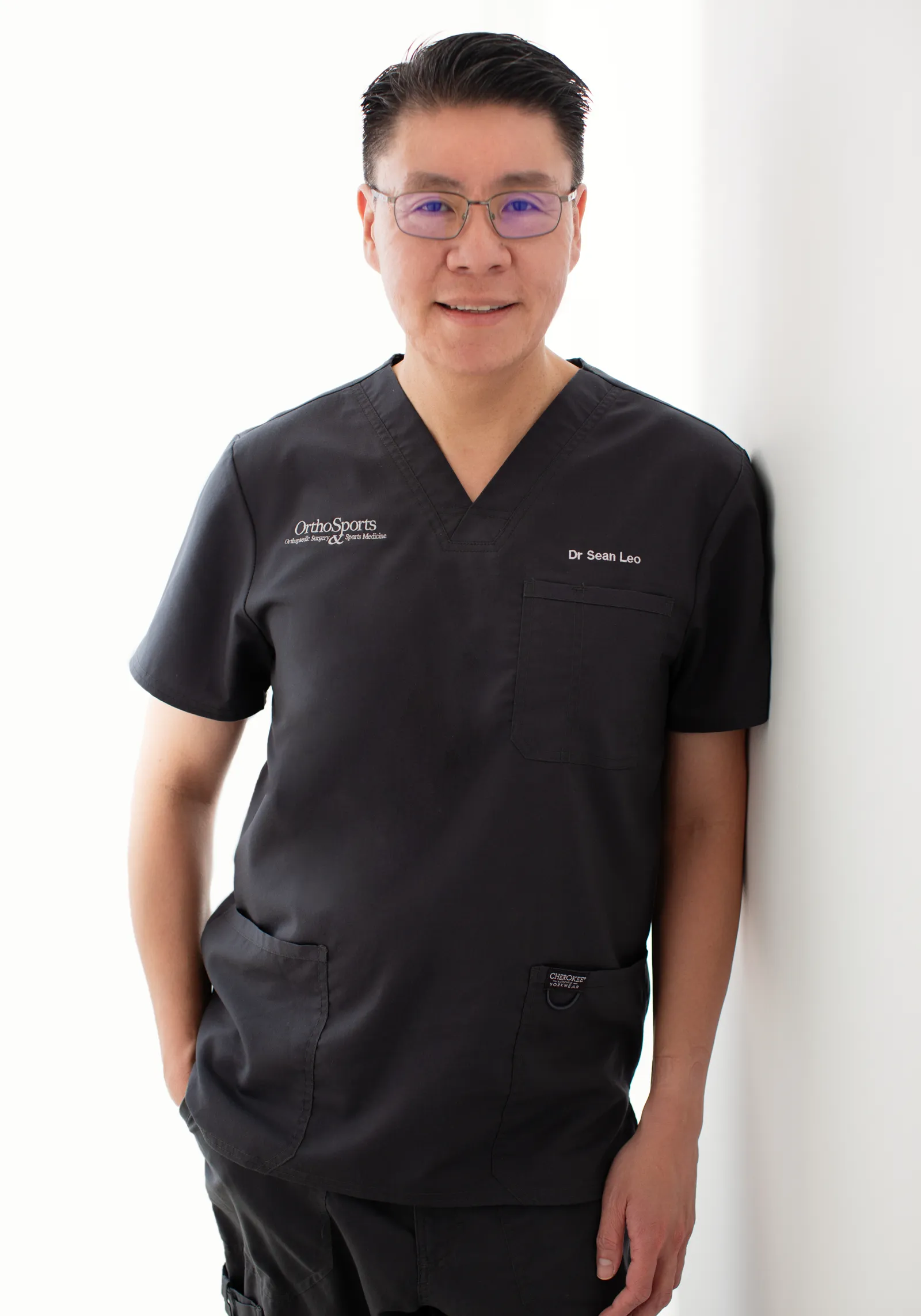
Meet Dr Sean Leo
Meet Dr Sean Leo
A consultant orthopaedic surgeon whose clinical focus is the knee, sports injuries and the lower limb.
- Dr Sean Leo is a consultant orthopaedic surgeon. OrthoKinetics is his professional brand; clinical services are provided through OrthoSports Orthopaedic & Sports Surgery at Mount Elizabeth Novena Specialist Centre and Connexion, Farrer Park Hospital.
- His clinical interests include sports injuries, including those in adolescent athletes; ligament reconstruction; realignment surgery; cartilage repair and joint-preservation approaches; knee replacement; and technology-assisted hip and knee surgery.
- The consistent aim is to return people to meaningful function and activity while avoiding unnecessary downtime.
- Qualifications: MBBS, MRCS (Ed), MMed (Ortho), FRCSEd (Ortho).
More detail: Meet Dr Sean Leo
- Training and career: Dr Leo trained in medicine at the National University of Singapore and served in the Singapore Armed Forces Medical Corps from 2000 to 2012, where his work included the care of soldiers in physically demanding roles. He completed postgraduate orthopaedic training in 2009 and a one-year clinical fellowship at Royal North Shore Hospital in Sydney, Australia, before joining Changi General Hospital as an orthopaedic consultant in 2012, where he later held the positions of Senior Consultant and Director of the Hip and Knee Service. With seniority, his educational contribution has evolved towards mentorship and support for junior doctors.
What should I do next?
What should I do next?
What should I do next?
Because the two problems overlap, an assessment is the sensible next step. It brings together your story, an examination and the imaging that helps. An appointment supports an informed decision. It is not a commitment to surgery.
- Meniscus treatment and the repair-or-trim decision: meniscus repair surgery.
- Cartilage procedures and recovery: cartilage repair surgery.
- For cartilage on its own, see knee cartilage damage: causes, symptoms and treatment.
- For timelines, see meniscus tear recovery time.
- Browse the Knee Hub for other knee problems.
- This page provides general information and cannot diagnose an individual condition. OrthoKinetics does not provide emergency or urgent care through this webpage or its WhatsApp contact.
Sources
References
References
- Fox AJS, et al. The human meniscus. Clin Anat. 2015. PMID:25125315.
- American Academy of Orthopaedic Surgeons. OrthoInfo: Articular Cartilage Restoration (orthoinfo.org); American Orthopaedic Society for Sports Medicine. Articular Cartilage Injuries (sportsmed.org).
- Kopf S, et al. The 2019 ESSKA meniscus consensus. Knee Surg Sports Traumatol Arthrosc. 2020. PMID:32052121. PMCID:PMC7148286.
- Beaufils P, et al. The 2016 ESSKA consensus on degenerative meniscus lesions. Knee Surg Sports Traumatol Arthrosc. 2017. PMID:28210788. PMCID:PMC5331096.
- Duong V, et al. Evaluation and treatment of knee pain: a review. JAMA. 2023. PMID:37874571.
- Siemieniuk RAC, Harris IA, Agoritsas T, et al. Arthroscopic surgery for degenerative knee arthritis and meniscal tears: a clinical practice guideline. BMJ. 2017;357:j1982. doi:10.1136/bmj.j1982. PMID:28490431.
- Smith BE, et al. Special tests for assessing meniscal tears within the knee. BMJ Evid Based Med. 2015. PMID:25724195.
- Stiell IG, et al. The Ottawa Knee Rule. Ann Emerg Med. 1995. PMID:7574120.
- Chen X, et al. Quantitative Magnetic Resonance Imaging Had Greater Sensitivity in Diagnosing Chondral Lesions of the Knee: A Systematic Review and Meta-analysis. Arthroscopy. 2024. PMID:38336108.
- National Institute for Health and Care Excellence. Osteoarthritis in over 16s: diagnosis and management (NG226). 2022. nice.org.uk/guidance/ng226.
- Katz JN, et al. Surgery versus physical therapy for a meniscal tear and osteoarthritis (MeTeOR). N Engl J Med. 2013. PMID:23506518.
- Sihvonen R, et al. Arthroscopic partial meniscectomy versus sham surgery for a degenerative meniscal tear (FIDELITY). N Engl J Med. 2013. PMID:24369076.
- O’Connor D, et al. Arthroscopic surgery for degenerative knee disease. Cochrane Database Syst Rev. 2022. PMID:35238404.
- Lee YS, et al. Return to sports after meniscal surgery. Clin J Sport Med. 2019. PMID:29023273.
- Bannuru RR, et al. OARSI guidelines for the non-surgical management of osteoarthritis. Osteoarthritis Cartilage. 2019. PMID:31278997.
- Prill R, et al. Formal EU-US Meniscus Rehabilitation 2024 Consensus, Part II: prevention, non-operative treatment, return to sport. Knee Surg Sports Traumatol Arthrosc. 2025;33(8):3014-3024. doi:10.1002/ksa.12689. PMID:40501314.
- Kalske R, Sihvonen R, Paavola M, et al. Arthroscopic Partial Meniscectomy for Degenerative Tear — 10-Year Outcomes. N Engl J Med. 2026;394(17):1757–1759. doi:10.1056/NEJMc2516079. PMID:42054686.
- Nassar JE, et al. Systematic review and meta-analysis of ACI, MACI, OAT and OCA. Knee Surg Sports Traumatol Arthrosc. 2025;33(8):2745-2762. doi:10.1002/ksa.12525. PMID:39497425. PMCID:PMC12310097.
Was this page helpful?