Introduction
Foot and ankle care · Achilles tendon
Achilles Tendon Rupture: assessment, treatment and recovery
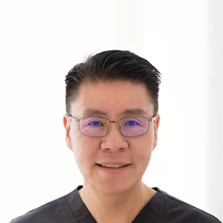
Dr Sean LeoOrthopaedic Surgeon, Singapore
Clinic enquiries · Not an emergency service
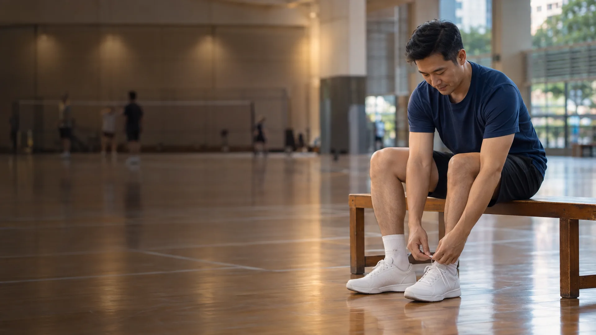
Illustrative image

Heard a “pop” in your heel?
Heard a “pop” in your heel?
Heard a “pop” in your heel?
- A sudden “pop”, followed by difficulty pushing off or rising onto your toes, is the classic story of an Achilles tendon rupture — an acute tear of the large tendon joining the calf muscle to the heel bone.
- This is an acute injury worth assessing promptly. Confirming what has torn, and how much, keeps your options open.
- Both non-operative functional rehabilitation and surgical repair are real treatment options with genuine trade-offs — this page sets out both, honestly, so the decision can be an individual one.
- OrthoKinetics is Dr Sean Leo’s professional brand. Clinical care is provided through OrthoSports Orthopaedic & Sports Surgery.
What’s covered on this webpage
Follow the route in order, or jump to the stop you need. Stops you have read are marked.
What an Achilles tendon rupture is
What an Achilles tendon rupture is
What an Achilles tendon rupture is
The Achilles tendon is a continuation of the calf muscles, attaching at the back of the heel bone. Because the calf muscles are strong and used repetitively, it’s the largest tendon in the body. A rupture is a tear in that tendon, and tears can be complete or partial — partial tears are rare compared with complete ones.
A complete tear separates the tendon into two ends, so the calf can no longer transmit force through it: without the tendon in one piece, you may not be able to lift your body up onto your toes on the injured side. That loss of push-off, more than the tear itself, is usually what brings people in for assessment.

How a rupture usually happens and how it feels
How a rupture usually happens and how it feels
How a rupture usually happens and how it feels
A very strong force is needed to tear the Achilles tendon — typically, tears happen when the calf muscle is stretched too quickly, during sudden push-off, sprinting or a rapid change of direction. Badminton, tennis and running are the classic Singapore recreational contexts for this kind of injury.
When the tendon tears, you may hear a popping noise, followed by severe pain and swelling near the heel; walking on the injured leg can become difficult, and standing up on the toes of that leg is often impossible.
Two things that raise the risk of a tear
A history of corticosteroid injections for Achilles tendinitis has been linked to an increased risk of rupture. Certain antibiotics — ciprofloxacin and levofloxacin among them — have also been linked with tendon tearing. Neither is a diagnosis on its own; they’re useful context if they apply to you.
Symptoms alone don’t confirm the diagnosis — they overlap with other calf and heel injuries, which is why an assessment matters, not just the story.

Is this a gradual overuse problem instead
Is this a gradual overuse problem instead
Is this a gradual overuse problem instead?
If your pain built up gradually with activity over weeks, without a sudden “pop” or snap, this page probably isn’t the right one. That pattern is more likely the chronic, overuse presentation — Achilles tendinitis — which is covered separately: Achilles tendonitis.
Suspected rupture
Suspected rupture
Suspected rupture: seek prompt assessment
A suspected Achilles rupture should be assessed promptly, so the injury can be confirmed and treatment options discussed while the window for either path is open. This webpage is not an emergency service. Severe trauma, an open wound, or signs of a clot or vascular problem (such as sudden calf swelling, pain or breathlessness) need an appropriate urgent-care route, not a routine website appointment. No list on this page can rule out a serious problem remotely.

How a rupture is confirmed
How a rupture is confirmed
How a rupture is confirmed
Diagnosis starts with the story of the injury and a physical examination. The main clinical test is the calf-squeeze test (also called the Thompson or Simmonds test): the clinician squeezes the calf and watches how the foot responds. In a prospective study of 174 patients with a confirmed tear, the calf-squeeze test was highly sensitive (0.96) at detecting a complete tear, and in a further 28 patients without a tear, highly specific (0.93) at correctly showing the tendon was intact. In plain terms: it’s a strong, reliable clue, particularly when the foot fails to flex on squeezing — but it’s one part of the picture, not used alone.
More on imaging
Ultrasound can show the tendon in real time and how it behaves as the ankle moves. Magnetic resonance imaging (MRI) gives a clearer picture of soft tissue than an X-ray, and can show the amount of tendon torn, its location, and help rule out a different injury with similar symptoms. Imaging is supportive, not mandatory — not every rupture needs an MRI, and a scan on its own doesn’t decide whether surgery is needed.
Structure / Patient / Goal
Structure / Patient / Goal
Deciding together
Structure / Patient / Goal (How options are weighed)
S
Structure
What is affected — whether the tear is complete or partial, its location, and how far the torn ends have pulled apart.
P
Patient
How it affects you — your age, activity level, general health (including whether you smoke), preferences, and how you feel about surgery versus a longer period of immobilisation.
G
Goal
What you hope to return to — walking comfortably, returning to work, running, or a pivoting sport such as badminton or tennis.
Your plan
No single factor — the tear grade, your age, or which sport you play — automatically decides the plan. These three considerations are weighed together.

The two management paths
The two management paths
The two management paths
Both paths share the same underlying aim: restoring the tendon’s ability to transmit force, safely. Both require a long, structured rehabilitation — neither is a quick fix, and neither is “doing nothing.”
Non-operative functional rehabilitation
This means protecting the tendon in a boot, then progressively restoring movement and loading it, under guidance, without surgery. It has become a legitimate first-line option as rehabilitation protocols have improved: in a multicentre randomised trial where both operative and non-operative patients received the same accelerated functional rehabilitation, there was no clinically important difference between the groups in strength, range of motion, calf circumference or functional score — and re-rupture was low in both arms (2 of 72 in the operative group, 3 of 72 in the non-operative group). A separate randomised trial using controlled early motion in both groups found the same pattern: comparable functional results and a low re-rupture rate in each, suggesting that controlled early motion — not surgery itself — is the important part of treatment.
Surgical repair
This means physically repairing the torn tendon ends, followed by the same kind of protected, progressive rehabilitation. Surgery’s advantage, where it exists, is on re-rupture; its cost is a real increase in wound, infection and nerve-related problems (covered honestly in Who should think carefully before surgery).
The re-rupture comparison
This is the comparison most competitor pages don’t make, so it’s worth stating plainly and with its caveats intact.
The largest and most recent pooled analysis (29 studies, nearly 16,000 patients) found a modest absolute reduction in re-rupture with surgery — about 2.3% after operative treatment versus about 3.9% after non-operative treatment (a risk difference of 1.6 percentage points; risk ratio 0.43, 95% confidence interval 0.31 to 0.60). But operative treatment also carried a significantly higher rate of *other* complications — about 4.9% versus 1.6% — driven mainly by infection. And critically: in the subset of studies that used accelerated functional rehabilitation with early range of motion, there was no significant difference in re-rupture rate between operative and non-operative treatment at all (risk ratio 0.60, 95% CI 0.26 to 1.37).
A separate meta-analysis of randomised trials found the same pattern from a different angle: where functional rehabilitation with early motion was used, re-rupture rates were equal between surgical and non-surgical patients (risk difference 1.7%, not statistically significant). Where it wasn’t used, surgery’s re-rupture advantage was larger — but came with a substantially higher rate of complications other than re-rupture (an absolute increase of 15.8%, favouring non-operative care on that measure).
The evidence over time
Older pooled trial data (12 randomised trials, 2005) found a bigger surgical advantage on re-rupture (relative risk 0.27, 95% CI 0.11–0.64) alongside a much higher complication risk (relative risk 10.60, 95% CI 4.82–23.28) — but these trials predate modern functional-rehabilitation protocols, and most of that “complication” figure reflects wound, adhesion and skin-sensation problems rather than anything more serious. A further quantitative review of randomised trials confirmed the same direction — lower re-rupture with surgery, but significantly more deep infection, non-cosmetic scarring and sural-nerve sensory disturbance.
In short: modern functional rehabilitation has narrowed the historical gap between the two paths considerably, and in some trial settings has closed it entirely. Surgery still carries, on the pooled evidence, a small absolute re-rupture advantage in some contexts, traded against a real increase in wound and infection-related problems. Neither path is free of risk, and neither is uniformly “better” — which is exactly why this is an individual decision.

What surgical repair involves — and cannot guarantee
What surgical repair involves — and cannot guarantee
What surgical repair involves — and cannot guarantee
The aim of repair is to restore the tendon’s continuity. If the tendon has torn away from its attachment at the heel, surgeons use anchor devices drilled into the heel bone; tears through the middle of the tendon are repaired with sutures bringing the torn edges together. If too much time has passed since the injury, the tendon can shorten and scar, and surgeons sometimes add donated tissue (allograft) to bridge the gap.
Repair restores continuity — it does not restore a risk-free tendon or guarantee a return to your previous level of sport. Some loss of strength is common after an Achilles rupture regardless of which path is taken; with focused strengthening and therapy, most people return to their previous level of activity, but this takes time (see Recovery).
It’s also worth being honest about the strength of the guideline evidence behind surgical technique: a professional clinical-practice guideline reviewing the evidence graded most of its recommendations as inconclusive or weak, with only two graded as moderate strength — both concerning early, protected weight-bearing and mobilisation after surgery, rather than the surgical technique itself.
Minimally invasive versus open repair
Minimally invasive versus open repair
Minimally invasive versus open repair
Where the evidence conflicts here, it’s presented as a conflict rather than resolved in favour of one technique.
A systematic review of minimally invasive (percutaneous) versus open repair found minimally invasive surgery had fewer complications overall, and an older meta-analysis of randomised trials similarly found a lower complication rate with percutaneous repair. But a larger, more recent comparative series (615 patients across three trauma centres) found no significant difference in overall complication rate between open and minimally invasive approaches (11.6% versus 13.2%).
What is consistent across the evidence is the *type* of trade-off: minimally invasive techniques tend to reduce wound-related problems, but carry a higher rate of sural-nerve sensory disturbance (a patch of numbness or altered sensation, typically on the outer side of the foot). The two approaches are broadly comparable in outcome, with a different complication profile rather than one being clearly safer overall.
Who should think carefully before surgery
Who should think carefully before surgery
Who should think carefully before surgery
Both paths carry real risks, and personal factors genuinely change the balance.
Risks of surgery include wound problems and infection (the pooled operative complication rate is around 4.9%, of which infection accounts for roughly 2.8%), sural-nerve sensory disturbance and non-cosmetic scarring, nerve injury more broadly, complications from anaesthesia, and — despite repair — the possibility of a further tear.
Risks of non-operative care centre on re-rupture, at a broadly similar order of magnitude to surgery once modern functional rehabilitation is used (see the re-rupture comparison above).
Personal factors that matter: in a series of 615 patients undergoing repair, active tobacco use was associated with a substantially higher complication risk (roughly three times higher), and advancing age was also independently associated with more complications. These are genuinely part of the decision, not afterthoughts — if you smoke, this is worth raising directly with your surgeon.
Deep-vein thrombosis (DVT) is common after an Achilles rupture, whichever path is chosen. In one screened study, about a third of patients had ultrasound evidence of a clot at eight weeks, with no significant difference between surgical and non-surgical treatment — often without symptoms. Be aware of calf pain, swelling, warmth or breathlessness during your recovery, and seek prompt medical attention if these occur. That judgement is made with your treating clinician.
Recovery and returning to activity
Recovery and returning to activity
Recovery
Recovery and returning to activity
Recovery from an Achilles rupture is long, and it’s staged rather than governed by a calendar. Typically, that means a period of immobilisation in a boot of at least around six weeks, followed by a gradual reduction in boot use as your doctor or therapist allows more range of motion, with strengthening exercises added progressively to rebuild push-off strength.
Full recovery commonly takes around 12 months; some people find it takes closer to two years to reach all of their goals. Some loss of strength is common regardless of which path was taken, and getting back to your previous level of activity — where that’s achieved — can take up to 18 to 24 months.
On return to sport specifically: a large systematic review pooling 108 studies and over 6,500 patients found that, overall, about 80% of people return to play after an Achilles rupture (95% confidence interval 75% to 85%). It’s worth reading that figure with its own caveat — studies that used a clearly defined way of measuring “return to play” reported meaningfully lower rates than studies without one, and “returning to play” is not the same thing as returning at your exact pre-injury level, which this evidence doesn’t separately quantify.

Recovery Reflection
Recovery reflection
If both paths carry real trade-offs, what actually makes one right for you?
It’s tempting to look for the objectively “better” option — the one with the lower number attached. But the honest evidence here doesn’t hand you a single winner: surgery’s small re-rupture advantage, where it exists, is traded against a real increase in wound and infection-related problems, and modern rehabilitation narrows even that gap. The right answer usually has less to do with which number is smaller and more to do with what you’re weighing it against — your tissue, your health, and what you’re hoping to get back to.
What does it mean to measure progress by what your tendon can do, rather than by the calendar?
A rupture asks for patience in a way that’s genuinely uncomfortable — there’s no shortcut past the months of protected loading and rebuilt strength. Watching for milestones — can you stand on your toes, can you push off evenly, can you load the tendon without it complaining — tends to be a steadier guide than counting weeks, and it keeps the goalposts realistic rather than arbitrary.
The milestones that matter here are what the tendon can do, such as standing on your toes, pushing off evenly and taking load without complaint, and they are agreed with your team, not dictated by a number of weeks.
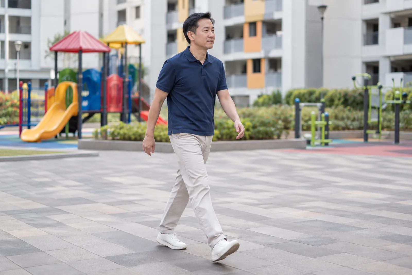
Illustrative image

 With you through the seasons
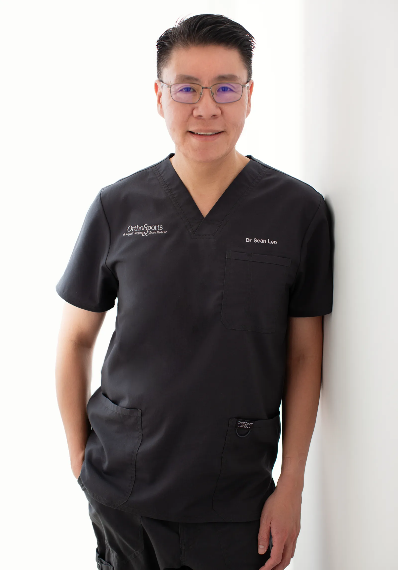
Meet Dr Sean Leo
Meet Dr Sean Leo
A consultant orthopaedic surgeon whose clinical focus is the knee, sports injuries and the lower limb.
- Dr Sean Leo is a consultant orthopaedic surgeon. OrthoKinetics is his professional brand; clinical services are provided through OrthoSports Orthopaedic & Sports Surgery at Mount Elizabeth Novena Specialist Centre and Connexion, Farrer Park Hospital.
- His clinical interests include sports injuries, including those in adolescent athletes; ligament reconstruction; realignment surgery; cartilage repair and joint-preservation approaches; knee replacement; and technology-assisted hip and knee surgery.
- The consistent aim is to return people to meaningful function and activity while avoiding unnecessary downtime.
- Qualifications: MBBS, MRCS (Ed), MMed (Ortho), FRCSEd (Ortho).
More detail: Meet Dr Sean Leo
- Training and career: Dr Leo trained in medicine at the National University of Singapore and served in the Singapore Armed Forces Medical Corps from 2000 to 2012, where his work included the care of soldiers in physically demanding roles. He completed postgraduate orthopaedic training in 2009 and a one-year clinical fellowship at Royal North Shore Hospital in Sydney, Australia, before joining Changi General Hospital as an orthopaedic consultant in 2012, where he later held the positions of Senior Consultant and Director of the Hip and Knee Service. With seniority, his educational contribution has evolved towards mentorship and support for junior doctors.
Questions people often ask
Common Achilles rupture questions
Common Achilles rupture questions
Do I need surgery for a torn Achilles?
Not automatically. Non-operative functional rehabilitation is a legitimate option, particularly where accelerated rehabilitation protocols are used, and modern evidence shows the re-rupture gap between the two paths has narrowed and in some studies closed entirely.
Surgery carries a small absolute re-rupture advantage in some studies (around 1.6 percentage points, pooled), but this comes with a higher rate of other complications — mainly wound and infection problems. Where accelerated functional rehabilitation with early controlled motion is used in both groups, trials have found no clinically important difference in outcome and no significant re-rupture difference. The decision depends on Structure, Patient and Goal, not a single rule.
Can a rupture heal without surgery?
Yes. With protected immobilisation followed by progressive, guided loading, non-operative treatment can produce comparable functional outcomes to surgery, with a similarly low re-rupture rate where accelerated rehabilitation is used.
A multicentre randomised trial giving both surgical and non-surgical patients the same accelerated functional rehabilitation found no clinically important difference in strength, movement, calf circumference or function, with low re-rupture in both groups (2 of 72 operative, 3 of 72 non-operative). This doesn’t mean every rupture should be treated non-operatively — that’s an individual decision — but it does mean non-operative care is a genuine option, not a fallback.
How is an Achilles rupture diagnosed?
Through a combination of the story of the injury, a physical examination — including the calf-squeeze (Thompson/Simmonds) test — and, where useful, imaging such as ultrasound or MRI.
The calf-squeeze test is highly sensitive and specific for a complete tear in the published diagnostic-accuracy evidence. Imaging supports rather than replaces this clinical picture: ultrasound shows the tendon moving in real time, and MRI can show the extent and location of a tear and help rule out other injuries. Not every rupture needs a scan.
What is the recovery time?
Recovery is measured in months, not weeks. Immobilisation typically runs at least around six weeks, with full recovery commonly taking around 12 months and, for some people, up to two years to reach all their goals.
Progress moves through stages — protected immobilisation, then progressive range of motion, then strengthening — guided by how the tendon is actually performing rather than a fixed date. Some strength loss is common regardless of treatment path.
Can I walk on a torn Achilles?
Immediately after a complete tear, walking normally and pushing off are usually not possible — that loss of push-off is part of what prompts assessment. Protected weight-bearing typically begins under guidance as part of a structured rehabilitation plan, whichever treatment path is chosen.
A complete tear means the calf can no longer transmit force through the tendon, so standing on your toes on the injured side is usually not possible before treatment. Both non-operative and surgical paths involve a period of protection followed by progressively increasing, clinician-guided weight-bearing — not unrestricted walking from day one.
What is the re-rupture risk?
Re-rupture is uncommon on either path. Pooled estimates put it at around 2.3% after surgery versus around 3.9% after non-operative treatment, though this gap narrows — and in some trial settings disappears — when accelerated functional rehabilitation is used.
The largest recent meta-analysis (nearly 16,000 patients) found a modest absolute difference of 1.6 percentage points favouring surgery overall, but no significant difference specifically in the subset of studies using accelerated functional rehabilitation with early motion. A separate randomised-trial meta-analysis found the same pattern: equal re-rupture rates under functional rehabilitation, a larger surgical advantage without it — offset by a higher rate of other complications.
When can I play sport again?
About 80% of people return to some level of sport after an Achilles rupture, though the exact figure depends on how “return to play” is measured, and this is not the same as returning at your exact pre-injury level.
A systematic review pooling 108 studies and over 6,500 patients found an overall return-to-play rate of 80% (95% CI 75–85%), with lower rates reported in studies that used a clearly defined return-to-play metric. Return to sport is judged by milestones — strength, movement quality, confidence loading the tendon — rather than a calendar date, and it is the final and most demanding stage of recovery.
What are the risks of surgery?
The main risks are wound problems and infection, sural-nerve sensory disturbance, scarring, and — despite repair — the possibility of a further tear. Smoking and older age are both associated with a higher complication risk.
The pooled operative complication rate is around 4.9%, of which infection accounts for roughly 2.8%. Sural-nerve sensory disturbance and non-cosmetic scarring are significantly more common after surgery than after non-operative care. In a series of 615 surgical patients, active tobacco use was linked to around a three-fold higher complication risk, and advancing age was independently linked to more complications too.
Is minimally invasive repair safer than open surgery?
Not clearly. The evidence conflicts: some studies show fewer overall complications with minimally invasive repair, while a larger, more recent series found no significant difference in overall complication rate between the two approaches.
What is consistent is the type of trade-off, not a verdict: minimally invasive repair tends to reduce wound-related problems but carries a higher rate of sural-nerve sensory disturbance. The two approaches are broadly comparable overall, and the right choice depends on your specific tear and circumstances, not a blanket rule.
How do I decide between non-operative care and surgery?
The decision weighs Structure (the tear itself), Patient (you — your health, activity level and preferences) and Goal (what you’re hoping to return to) — not a single factor like age or tear grade alone.
No single feature decides the plan on its own. A complete tear does not automatically mean surgery; being younger or more active does not automatically mean surgery either. Structure, Patient and Goal are weighed together, alongside the honest trade-offs on re-rupture and complications set out above.
Costs and insurance
Costs and insurance
Practicalities
Cost, Medisave and insurance

A cost estimate for surgery can be given only after a consultation and assessment, once a surgical plan has been developed. Each case is unique — the treatment involved, time taken and other factors all contribute to the overall estimate. The clinic will provide a written estimate should you proceed with treatment, and this can also assist in obtaining pre-authorisation from insurance where applicable.
Insurance panels. Dr Sean Leo, Orthopaedic Surgeon, is on the specialist panels of Great Eastern (GE Panel), HSBC Life (HSBC Life Shield) and Singlife (Singlife Shield), and on the Prudential Panel. AXA Insurance Singapore became part of HSBC Life in 2023, and former AXA Shield plans are now HSBC Life Shield plans.
Corporate health plans. Dr Leo is on the panels of both Alliance Medinet and iXchange, which administer medical benefits for many company health plans. If your company plan uses either panel, you can see Dr Leo as a panel doctor.
If your company uses a different panel. Some company plans still let you see a doctor of your choice outside their panel, then claim reimbursement. This varies from plan to plan, as may the co-payment, so please check your benefits with your company’s HR department.
Panel arrangements change from time to time, so please confirm with your insurer or your company’s plan administrator before your appointment.
Dr Leo is on the panels shown. If your insurer is not listed, you may ask whether co-management with an Orthosports surgeon partner on that panel is clinically and administratively appropriate. Orthosports surgeon partners are collectively on the panels of all Shield plans.
Arrange a consultation: the appointment section is further down this page.
Sources
References
References
- American Academy of Orthopaedic Surgeons (AAOS). Achilles Tendon Rupture (Tear). OrthoInfo. Available at: https://orthoinfo.aaos.org/en/diseases–conditions/achilles-tendon-rupture-tear
- Maffulli N. The clinical diagnosis of subcutaneous tear of the Achilles tendon. A prospective study in 174 patients. Am J Sports Med. 1998;26(2):266–270. PMID 9548122.
- Ochen Y, Beks RB, van Heijl M, et al. Operative treatment versus nonoperative treatment of Achilles tendon ruptures: systematic review and meta-analysis. BMJ. 2019;364:k5120. doi:10.1136/bmj.k5120. PMID 30617123. PMC6322065. Open access.
- Soroceanu A, Sidhwa F, Aarabi S, Kaufman A, Glazebrook M. Surgical versus nonsurgical treatment of acute Achilles tendon rupture: a meta-analysis of randomized trials. J Bone Joint Surg Am. 2012;94(23):2136–2143. PMID 23224384. PMC3509775. Open access.
- Khan RJ, Fick D, Keogh A, Crawford J, Brammar T, Parker M. Treatment of acute Achilles tendon ruptures. A meta-analysis of randomized, controlled trials. J Bone Joint Surg Am. 2005;87(10):2202–2210. PMID 16203884.
- Willits K, Amendola A, Bryant D, et al. Operative versus nonoperative treatment of acute Achilles tendon ruptures: a multicenter randomized trial using accelerated functional rehabilitation. J Bone Joint Surg Am. 2010;92(17):2767–2775. PMID 21037028.
- Twaddle BC, Poon P. Early motion for Achilles tendon ruptures: is surgery important? A randomized, prospective study. Am J Sports Med. 2007;35(12):2033–2038. PMID 17885221.
- Wilkins R, Bisson LJ. Operative versus nonoperative management of acute Achilles tendon ruptures: a quantitative systematic review of randomized controlled trials. Am J Sports Med. 2012;40(9):2154–2160. PMID 22802271.
- Chiodo CP, Glazebrook M, Bluman EM, et al. Diagnosis and treatment of acute Achilles tendon rupture. J Am Acad Orthop Surg. 2010;18(8):503–510. PMID 20675643. (AAOS clinical-practice guideline.)
- Del Buono A, Volpin A, Maffulli N. Minimally invasive versus open surgery for acute Achilles tendon rupture: a systematic review. Br Med Bull. 2014;109:45–54. PMID 24126569.
- Stavenuiter XJR, Lubberts B, Prince RM 3rd, Johnson AH, DiGiovanni CW, Guss D. Postoperative complications following repair of acute Achilles tendon rupture. Foot Ankle Int. 2019;40(6):679–686. PMID 30808187.
- Nilsson-Helander K, Thurin A, Karlsson J, Eriksson BI. High incidence of deep venous thrombosis after Achilles tendon rupture: a prospective study. Knee Surg Sports Traumatol Arthrosc. 2009;17(10):1234–1238. PMID 19238360.
- Zellers JA, Carmont MR, Grävare Silbernagel K. Return to play post-Achilles tendon rupture: a systematic review and meta-analysis of rate and measures of return to play. Br J Sports Med. 2016;50(21):1325–1332. PMID 27259751. PMC5136353. Open access.
Final next step
Final next step
Next step
Final next step
Reading about a problem is a starting point. An assessment brings together the history, an examination where needed and any useful tests, and helps you and your clinician decide what to do next. How much physical examination is needed is a matter of clinical judgement. It may be limited when pain makes an examination unreliable, or when imaging clearly shows a problem that matches the symptoms, such as a fracture seen on an X-ray or an MRI scan that clearly shows an injury consistent with the symptoms. The Singapore Medical Council and the UK General Medical Council both allow remote medical consultations, where a physical examination is not possible, with safeguards: the doctor must be clear about the limits of an opinion formed without an examination, and whether an in-person examination is needed, or referral to other services, is left to the clinical judgement of the attending doctor.
Signs that need urgent help
Some symptoms should not wait for a routine appointment. Each item below says where to go.
After an injury
- The ankle or foot sits at an odd angle, a bone is showing, there is a deep cut, you are in severe pain, or the toes look blue or white or feel numb. Go to an emergency department or call 995.
- You felt a sudden pop or sharp pain at the back of the heel or calf, and pushing off or rising onto your toes is difficult. Arrange an urgent same-day assessment.
- It is very painful or the pain is getting worse, the swelling or bruising is large or spreading, or it hurts to put weight on the ankle or foot. Arrange an urgent same-day assessment.
At any time
- A joint, such as the ankle, is suddenly very painful, hot, red and swollen, especially with a fever or feeling generally unwell. Go to an emergency department or call 995.
- One calf or thigh is newly painful and swollen. Arrange an urgent same-day assessment. If you also feel short of breath or have chest pain, go to an emergency department or call 995.
If you have diabetes
- You have a foot sore and a fever or feel very unwell, or the foot is cold, pale or blue. Or skin on the foot or toes has turned black, or a sore looks deep, or there is pus or redness spreading up the foot. Go to an emergency department or call 995.
- You have a new foot sore, or a foot that is hot, swollen, changed in colour or leaking fluid, without the signs above. Arrange an urgent assessment within 24 hours.
If you are admitted by the doctor at the urgent care centre of one of these private hospitals, the Parkway Pantai hospitals (Mount Elizabeth Orchard, Mount Elizabeth Novena, Gleneagles and Parkway East), Farrer Park Hospital and Thomson Medical Centre, you may ask for Dr Sean Leo to be your attending orthopaedic surgeon. If you are instead discharged and referred for orthopaedic follow-up, you may contact the clinic to arrange an appointment. Both are subject to the hospital’s admission processes and to Dr Leo’s availability.
Was this page helpful?
Arrange a consultation
Appointments are arranged through the OrthoSports clinic team. Send your details below, or message or call the clinic.
Leave your details and the clinic team will reply to arrange a time.
Message the clinic on WhatsApp
-
38 Irrawaddy Road, #10-41 Mount Elizabeth Novena Specialist Centre, Singapore 329563 Map for Orthosports@Novena (opens Google Maps)
-
1 Farrer Park Station Road, #15-17 Connexion, Singapore 217562 Map for Orthosports@Connexion (opens Google Maps)
WhatsApp is for clinic enquiries only. It is not an emergency service and is not monitored for urgent care. Website or messaging contact is not an emergency-response service. For a medical emergency, use your local emergency service.
- OrthoKinetics does not provide emergency or urgent care through this webpage or its WhatsApp contact.