
Introduction
Foot and ankle care · Heel pain
Plantar fasciitis and heel pain: assessment and treatment

Dr Sean LeoOrthopaedic Surgeon, Singapore
Clinic enquiries · Not an emergency service
Illustrative image

Most plantar heel pain settles without surgery.
Most plantar heel pain settles without surgery.
Most plantar heel pain settles without surgery.
Plantar fasciitis is irritation and degeneration of the strong band under the foot (the plantar fascia) where it attaches to the heel. It is the most common cause of pain under the heel. It usually settles with stretching, footwear changes and load changes over months. Surgery is rarely needed.[1][2][4]
Heel symptoms overlap. This page helps you describe what you feel and understand how it is assessed. It cannot confirm what is causing your pain.
Seeing a specialist about a foot or ankle problem does not mean surgery has already been decided. An assessment helps you understand the problem and weigh reasonable options.
OrthoKinetics is Dr Sean Leo’s professional brand and patient-education platform. Clinical consultations and treatment are provided through OrthoSports Orthopaedic & Sports Surgery. Dr Sean Leo is a specialist orthopaedic surgeon who treats foot and ankle problems.
What’s covered on this webpage
Follow the route in order, or jump to the stop you need. Stops you have read are marked.
What the plantar fascia is and what it does
What the plantar fascia is and what it does
What the plantar fascia is and what it does
The plantar fascia is a very dense, tough fibre. It attaches the front part of the foot to the heel bone.
It runs along the sole and supports the arch. It takes load with every step. The point where it joins the heel bone carries a lot of that load, and that is where plantar fasciitis hurts.
Doctors may also call the condition plantar fasciopathy. That name describes changes in the fibres more than simple inflammation.[5]

How plantar fasciitis develops
How plantar fasciitis develops
How plantar fasciitis develops
It starts with load. Repeated strain where the fascia meets the heel can irritate the fibres and, over time, change them.[5]
Several things are linked with it:[2]
- a tight calf, with limited upward movement of the ankle;
- higher body weight, in people who are not athletes;
- running;
- work on your feet, especially on hard surfaces with poor shock absorption.
Flat feet and high-arched feet are also listed as risk factors.[10]
These are associations, not blame. In most cases, plantar fasciitis develops without a specific, identifiable reason.[9]

What plantar fasciitis feels like
What plantar fasciitis feels like
What plantar fasciitis feels like
People commonly describe this pattern:[2][9]
- sharp pain with the first steps after rest, most often first thing in the morning;
- pain at the inner, underside part of the heel;
- easing within a few minutes of walking;
- pain returning after long periods of standing, or late in the day.
The pain is often greater after activity than during it.[9]
This is the classic pattern. It is typical, not universal, and it describes symptoms without confirming a diagnosis. Other causes of heel pain can look different, and the next section covers them.
Heel pain is not always plantar fasciitis.
Heel pain is not always plantar fasciitis.
Heel pain is not always plantar fasciitis.
“Heel pain” is an umbrella term, and several problems sit under it.[2] Where the pain is gives a first clue.
| Where and how it hurts | What it may point to | Where to go |
|---|---|---|
| Under the heel, worst with the first steps after rest | The pattern of plantar fasciitis | Keep reading this page |
| At the back of the heel, building up gradually with activity | A different problem, such as Achilles tendonitis | See the Achilles tendonitis page |
| After a fall or impact; pain at rest or at night; numbness or tingling; swelling of both heels; fever or feeling generally unwell | Other causes | Arrange an assessment |
Other causes under the heel-pain umbrella include a heel fat-pad problem, a stress fracture, nerve irritation (such as tarsal tunnel syndrome) and tendon trouble where the Achilles meets the heel.[2][5]
Matching a row is a clue, not a diagnosis.
When should heel pain be assessed?
When should heel pain be assessed?
When should heel pain be assessed?
Plantar fasciitis is common and usually not serious.[4][5] Many people try sensible self-care first, and that is a reasonable place to start.
Heel pain is worth assessing when it continues despite that, keeps returning or limits ordinary activity.
Some features move assessment up the list. Arrange an assessment rather than waiting if heel pain:[2][5]
- follows a fall, an impact or another injury;
- is present at rest or at night;
- comes with numbness or tingling;
- involves swelling of both heels;
- comes with a fever or feeling generally unwell.
Safety note: This webpage is not an emergency service. The signs that need urgent help are listed at the end of this page. No list on this page can rule out a serious problem remotely.

How plantar fasciitis is assessed
How plantar fasciitis is assessed
How plantar fasciitis is assessed
Diagnosis starts with a conversation and an examination.[1][3]
Your clinician asks about the pain, your work and activity, your footwear and past treatment. The examination looks for tenderness at the inner heel and checks calf and ankle flexibility.[2][3]
The windlass test may be used. The examiner passively bends your big toe upwards at its base joint (the first metatarsophalangeal joint) to see whether this reproduces your heel pain. The test is positive if the pain is brought on, and a positive windlass test is one of the findings the guideline lists when diagnosing plantar fasciitis.[2][11]
Most people do not need a scan. Routine X-rays are not necessary for typical plantar fasciitis that has not followed an injury.[3]
When a scan helps
Imaging is useful when the picture is unusual, when a stress fracture is suspected, or when appropriate first treatment has not worked.[3] Ultrasound is a reasonable first option for pain that persists beyond about three months despite treatment.[4] A scan can look for other causes.
What about a heel spur? A spur on an X-ray report can sound alarming, but it does not change the diagnosis or the treatment.[3] Most people who have heel spurs do not have heel pain.[9] A spur can follow long-standing tension where the fascia attaches, so the video below calls it an after effect, not the cause.
Watch: What is a heel spur? (1 min 13 s)
Treatment starts with simple, sustained measures.
Treatment starts with simple, sustained measures.
Treatment starts with simple, sustained measures.
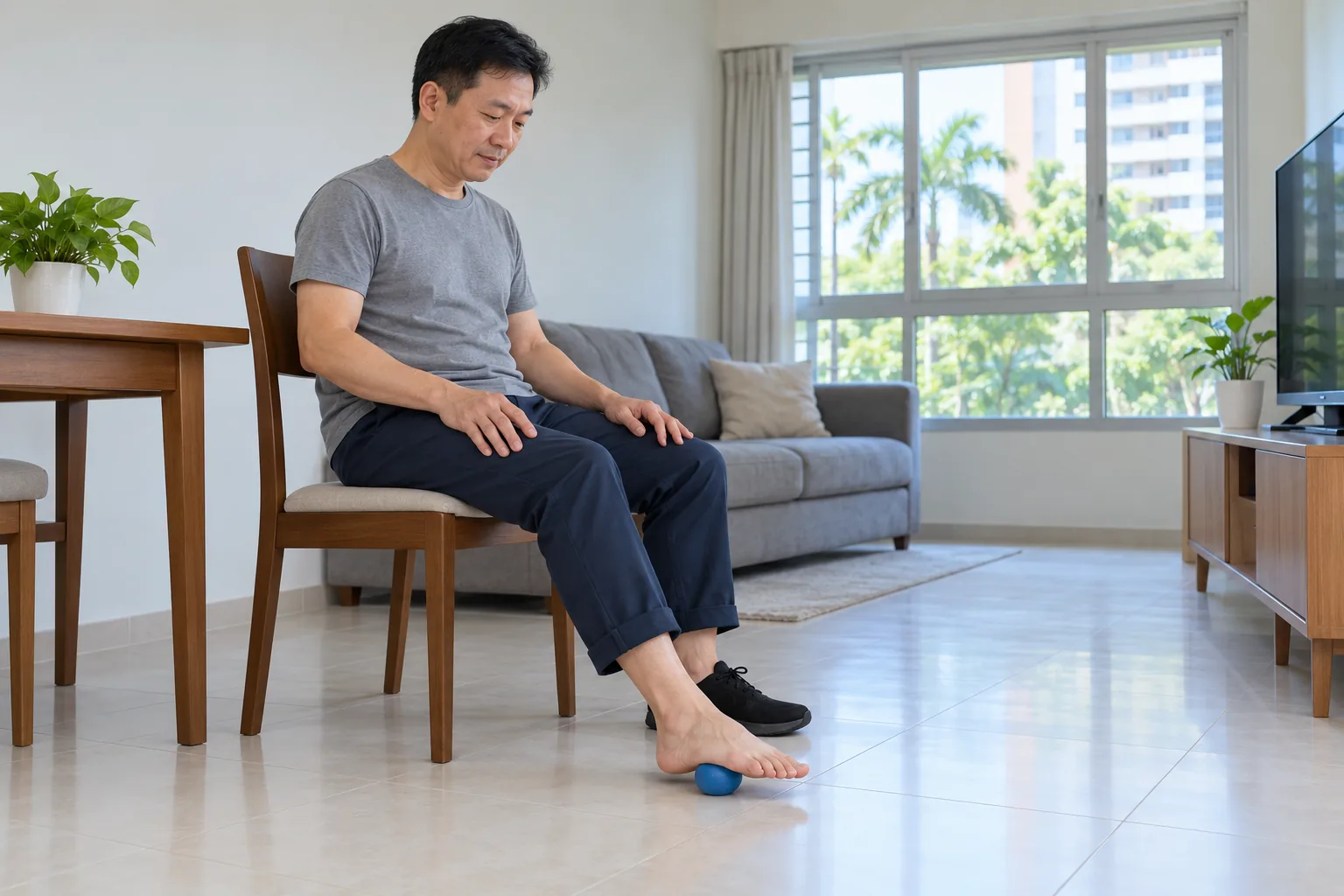
The first plan is a programme that you take part in. It usually combines:[1][2]
- an explanation of what is happening;
- changes to activity and load;
- stretching of the plantar fascia and the calf muscles;
- footwear guidance, with simple pain relief and ice massage when needed.
Stretching has the strongest evidence. Plantar fascia stretching and calf stretching (the gastrocnemius and soleus muscles) reduce pain and improve function in the short and long term. The 2023 US clinical practice guideline gives this its highest grade of recommendation.[2]
A physiotherapist usually guides the programme. You still do the exercises yourself.[10]
Calf flexibility, footwear and cushioning, body weight where relevant and the surfaces you stand on all change the load on the heel.[2][6] See what to look for in a shoe.
How long does it take? Improvement comes over weeks to months. About 80% to 90% of people improve within 12 months, and around 10% develop persistent symptoms.[4][5][6] These are group figures, not a promise for your heel.
Other measures that can help for a short time
- Foot taping, rigid or elastic, can help pain and function for short-term relief, alongside other physiotherapy.[2]
- Hands-on joint and soft-tissue treatment (manual therapy) also helps pain and function.[2]
- A night splint worn for one to three months may suit people whose pain is worst with the first step in the morning.[2]
These support the programme. They are not stand-alone cures.
Insoles and orthoses
Insoles (orthoses) are not recommended as the only short-term treatment. They may help when combined with other care.[2] In one trial, shop-bought and custom-made inserts did not differ for pain at 12 months.[5] A custom pair is therefore not proven better than a simple one.
The video below explains the types of insole. Which type suits you is a decision for your assessment. Watch: Orthotics: Insoles and Arch Supports Explained (1 min 58 s)
When is surgery considered?
When is surgery considered?
When is surgery considered?
Surgery is a last resort for a small minority. It is considered when disabling pain persists despite a full course of non-operative care.[4][5] Many months of a genuine trial of simpler care come first.
How long do sources suggest waiting?
Sources differ on the exact point. A Singapore consensus guideline says surgery may be offered when symptoms last more than six months and conservative treatment has failed. The guideline notes that the evidence behind this advice is limited, so it is a guide rather than a firm rule.[1] One review advises waiting about 12 months.[5]
Structure / Patient / Goal
Structure / Patient / Goal
Deciding together
Structure / Patient / Goal
S
Structure: what does the heel show?
Structure is the pattern of pain, where the heel is tender and how flexible the calf and ankle are. It includes what any scan added and which other causes were considered.
P
Patient: how does it affect you?
Patient is you as a whole person: your work, running, body weight, other health conditions, treatments genuinely tried, and how you feel about a plan that runs for months.
G
Goal: what are you hoping to do?
Goal is what matters to you, such as walking comfortably, working without a limp or returning to running. It shapes how long to persist and what to add next.
Your plan
No single factor decides the plan. Sticking with a programme is an active choice.
The decision is usually about persistence, not an operation.
The decision is usually about persistence, not an operation.
The decision is usually about persistence, not an operation.
Most plans need tuning before anything is added. The load, footwear or stretching may need adjusting. If progress stalls, the diagnosis is checked again.
Adding a treatment is part of the plan, not a sign of failure. Three options may come into the conversation.
Shockwave therapy
Extracorporeal shockwave therapy (ESWT) is a non-invasive treatment for persistent heel pain. A Singapore consensus guideline describes it as a safe and effective adjunct for defined indications.[1] It does not work for everyone.
What the research shows about shockwave and injection
A 2024 meta-analysis of 16 randomised trials (1,121 people) found shockwave reduced pain and improved function more than corticosteroid injection at 3 to 6 months, with no difference at 12 months. The authors note the 3-month result may be affected by publication bias. Radial and high-intensity protocols performed best in subgroup analyses.[8]
Corticosteroid injection brings relief that is usually short, about 4 to 12 weeks in randomised trials.[6] A separate 2018 meta-analysis cited in a family-medicine review found injection more effective than non-invasive treatments within three months.[4]
Corticosteroid injection
An injection can give meaningful relief, but it is short term.[6] It also carries risks. The plantar fascia can rupture. Rupture was reported in 2.4% to 6.7% of people in retrospective studies. The heel’s fat pad can also thin (fat-pad atrophy).[6] Repeated injections and higher body weight raise the rupture risk.[6]
A rupture can leave lasting problems. In one case series, 26 of 39 people were still symptomatic a year later.[7]
The Singapore guideline suggests injection only after conservative treatment has failed, placed around the fascia under ultrasound guidance. The guideline makes this a cautious suggestion because the pain relief is short-lived and the injection carries some risks.[1]
Surgery
Surgery is the rare endpoint. See When is surgery considered?.

What surgery for plantar fasciitis involves
What surgery for plantar fasciitis involves
What surgery for plantar fasciitis involves
Two operations are described. One releases part of the plantar fascia. The other, increasingly used, releases the calf muscle (gastrocnemius). Both are for the small group whose pain persists despite full non-operative care.[5]
Surgery has its own risks. The arch can lose some of its support (arch destabilisation), the middle of the foot can become strained and sore, and pain can continue. Around 5% to 10% of people still have pain after surgery.[4][5]
Recovery and return to activity
Recovery and return to activity
Recovery
Recovery and return to activity
Recovery is gradual and comes in steps over weeks to months. Flare-ups can happen when activity increases. About 80% to 90% of people improve within 12 months.[4][5]
Return to activity is judged by criteria, not by a calendar date. Walking without a limp, morning pain settling and tolerating the load you add are the signs your clinician looks for. Each step up in activity is judged by how the heel responds.

Risks and complications
Risks and complications
Risks and complications
Every path carries real limits.
- Persistent symptoms. Around 1 in 10 people develop persistent symptoms despite treatment.[6]
- Injection. Possible fascia rupture (2.4% to 6.7% in retrospective studies), fat-pad thinning, and lasting problems after a rupture.[6][7]
- Surgery. Arch destabilisation, midfoot strain, and continued pain in about 5% to 10%.[4][5]
- Passive fixes. Relying on repeated injections or costly insoles in place of the programme costs time and money, and injections add the risks above.
Does treatment work, and how long does it last
Does treatment work, and how long does it last
Does treatment work, and how long does it last?
Stretching and the first-line programme carry the strongest recommendations in the guidelines.[1][2] Shockwave therapy shows benefit at 3 to 6 months, with no proven difference from injection by one year.[8]
Most people improve. Symptoms can come back when your load changes. If they do, the same approach is used again.

OrthoKinetics Recovery Reflection
Recovery reflection
What is it like when the heel takes months to settle?
Every morning begins with the same first steps. Slow progress can wear you down. That does not mean the plan is failing. Steady, plain care, done most days, is what most heels respond to.
A heel that improves slowly is still improving.
Illustrative image

 With you through the seasons
Meet Dr Sean Leo
Meet Dr Sean Leo
A consultant orthopaedic surgeon whose clinical focus is the knee, sports injuries and the lower limb.
- Dr Sean Leo is a consultant orthopaedic surgeon. OrthoKinetics is his professional brand; clinical services are provided through OrthoSports Orthopaedic & Sports Surgery at Mount Elizabeth Novena Specialist Centre and Connexion, Farrer Park Hospital.
- His clinical interests include sports injuries, including those in adolescent athletes; ligament reconstruction; realignment surgery; cartilage repair and joint-preservation approaches; knee replacement; and technology-assisted hip and knee surgery.
- The consistent aim is to return people to meaningful function and activity while avoiding unnecessary downtime.
- Qualifications: MBBS, MRCS (Ed), MMed (Ortho), FRCSEd (Ortho).
More detail: Meet Dr Sean Leo
- Training and career: Dr Leo trained in medicine at the National University of Singapore and served in the Singapore Armed Forces Medical Corps from 2000 to 2012, where his work included the care of soldiers in physically demanding roles. He completed postgraduate orthopaedic training in 2009 and a one-year clinical fellowship at Royal North Shore Hospital in Sydney, Australia, before joining Changi General Hospital as an orthopaedic consultant in 2012, where he later held the positions of Senior Consultant and Director of the Hip and Knee Service. With seniority, his educational contribution has evolved towards mentorship and support for junior doctors.

Questions people often ask
Common plantar fasciitis questions
Common plantar fasciitis questions
Why does my heel hurt first thing in the morning?
Plantar fasciitis typically causes sharp pain with the first steps after rest. It usually eases within minutes of walking.[2][9] An assessment confirms the cause.
Can plantar fasciitis go away on its own?
Many cases settle with time and simple care. About 80% to 90% of people improve within 12 months.[4][5] Stretching is advised meanwhile.[1][2]
How long does it take to settle?
Weeks to months, and your own course varies. Around 10% of people develop persistent symptoms.[6]
Is a heel spur causing my pain?
Usually not. A spur does not change the diagnosis or the treatment.[3] The heel spur video explains more.
What is the difference between plantar fasciitis and Achilles tendonitis?
Plantar fasciitis hurts under the heel, worst with the first steps. Achilles tendonitis hurts at the back of the heel and builds with activity. See the Achilles tendonitis page.
Can insoles help?
They may help alongside other care. Custom pairs are not proven better than simple ones.[2][5]
What is shockwave therapy?
Extracorporeal shockwave therapy (ESWT) is a non-invasive option for persistent heel pain. It does not work for everyone.[1][8]
When is an injection or surgery considered?
After simpler care has not worked. Injection gives short relief with a small risk of rupture. Surgery is rare.[1][5][6]
Can it come back?
It can, when your load changes. The same approach is used again.
Costs and insurance
Costs and insurance
Practicalities
Cost, MediSave and insurance

Ask the clinic team for current information about fees, panel status, pre-authorisation and expected payment arrangements. Insurance coverage and out-of-pocket costs depend on the individual policy and the insurer’s decision; no coverage is guaranteed.
A cost estimate for surgery can be given only after a consultation and assessment, once a surgical plan has been developed. Each individual condition is unique — implants used, time taken, and other factors contribute to the overall estimate. The estimate will be provided to you by the clinic should you decide on surgical management, and will also be used to assist in obtaining pre-authorisation from insurance where applicable.
A cost estimate for surgery can be given only after a consultation and assessment, once a surgical plan has been developed. A surgical estimate separates surgeon, anaesthetist, hospital or facility, implant and consumable costs, and the MOH publishes national fee benchmarks for common procedures. For how costs, MediSave and insurance work in Singapore, see Costs, insurance and MediSave, explained.
Insurance panels. Dr Sean Leo, Orthopaedic Surgeon, is on the specialist panels of Great Eastern (GE Panel), HSBC Life (HSBC Life Shield) and Singlife (Singlife Shield), and on the Prudential Panel. AXA Insurance Singapore became part of HSBC Life in 2023, and former AXA Shield plans are now HSBC Life Shield plans.
Corporate health plans. Dr Leo is on the panels of both Alliance Medinet and iXchange, which administer medical benefits for many company health plans. If your company plan uses either panel, you can see Dr Leo as a panel doctor.
If your company uses a different panel. Some company plans still let you see a doctor of your choice outside their panel, then claim reimbursement. This varies from plan to plan, as may the co-payment, so please check your benefits with your company’s HR department.
Panel arrangements change from time to time, so please confirm with your insurer or your company’s plan administrator before your appointment.
Dr Leo is on the panels shown. If your insurer is not listed, you may ask whether co-management with an Orthosports surgeon partner on that panel is clinically and administratively appropriate. Orthosports surgeon partners are collectively on the panels of all Shield plans.
Specific benefits, pre-authorisation, reimbursement and cashless treatment depend on the individual policy and insurer approval and are not guaranteed.
Ready to book? The appointment section is at the end of this page.
Arrange a consultation: the appointment section is further down this page.
Sources
References
References
- Tan VAK, Tan CC, Yeo NEM, Zhang M, Mehta KV, Tian RHH, Tan B. Consensus statements and guideline for the diagnosis and management of plantar fasciitis in Singapore. Ann Acad Med Singap. 2024;53(2):101-112. doi:10.47102/annals-acadmedsg.2023211. PMID 38920234.
- Heel Pain – Plantar Fasciitis: Revision 2023. Clinical practice guideline, Academy of Orthopaedic Physical Therapy, APTA. J Orthop Sports Phys Ther. 2023;53(12):CPG1-CPG39. doi:10.2519/jospt.2023.0303.
- American College of Foot and Ankle Surgeons. Clinical Consensus Statement: Diagnosis and Treatment of Adult Acquired Infracalcaneal Heel Pain. J Foot Ankle Surg. 2018;57:370-381. Available at: https://www.acfas.org/ACFAS/media/ACFAS_Media/Heel-Pain-CCS.pdf
- Plantar Fasciitis. Am Fam Physician. 2019 (15 June). Available at: https://www.aafp.org/afp/2019/0615/p744
- Plantar fasciopathy: a current concepts review. Available at: https://pmc.ncbi.nlm.nih.gov/articles/PMC6134886/
- The effectiveness of corticosteroid injection in the treatment of plantar fasciitis. Review of 10 randomised controlled trials. Available at: https://pmc.ncbi.nlm.nih.gov/articles/PMC4545130/
- Acevedo JI, Beskin JL. Complications of plantar fascia rupture associated with corticosteroid injection. Foot Ankle Int. 1998. PMID 9498581.
- Efficacy of extracorporeal shockwave therapy compared with corticosteroid injections in plantar fasciitis: systematic review and meta-analysis. 2024. PMID 38738305.
- American Academy of Orthopaedic Surgeons (AAOS). Plantar Fasciitis and Bone Spurs. OrthoInfo. Available at: https://orthoinfo.aaos.org/en/diseases–conditions/plantar-fasciitis-and-bone-spurs/
- Health Promotion Board, Singapore. Plantar Fasciitis. HealthHub. Available at: https://www.healthhub.sg/health-conditions/plantar-fasciitis
- Buchanan B, Sina R, Kushner D. Plantar Fasciitis. StatPearls [Internet]. Treasure Island (FL): StatPearls Publishing. NBK431073. Available at: https://www.ncbi.nlm.nih.gov/books/NBK431073/
Final next step
Final next step
Next step
Final next step
Reading about a problem is a starting point. An assessment brings together the history, an examination where needed and any useful tests, and helps you and your clinician decide what to do next. How much physical examination is needed is a matter of clinical judgement. It may be limited when pain makes an examination unreliable, or when imaging clearly shows a problem that matches the symptoms, such as a fracture seen on an X-ray or an MRI scan that clearly shows an injury consistent with the symptoms. The Singapore Medical Council and the UK General Medical Council both allow remote medical consultations, where a physical examination is not possible, with safeguards: the doctor must be clear about the limits of an opinion formed without an examination, and whether an in-person examination is needed, or referral to other services, is left to the clinical judgement of the attending doctor.
Signs that need urgent help
Some symptoms should not wait for a routine appointment. Each item below says where to go.
After an injury
- The ankle or foot sits at an odd angle, a bone is showing, there is a deep cut, you are in severe pain, or the toes look blue or white or feel numb. Go to an emergency department or call 995.
- You felt a sudden pop or sharp pain at the back of the heel or calf, and pushing off or rising onto your toes is difficult. Arrange an urgent same-day assessment.
- It is very painful or the pain is getting worse, the swelling or bruising is large or spreading, or it hurts to put weight on the ankle or foot. Arrange an urgent same-day assessment.
At any time
- A joint, such as the ankle, is suddenly very painful, hot, red and swollen, especially with a fever or feeling generally unwell. Go to an emergency department or call 995.
- One calf or thigh is newly painful and swollen. Arrange an urgent same-day assessment. If you also feel short of breath or have chest pain, call 995.
If you have diabetes
- You have a foot sore and a fever or feel very unwell, or the foot is cold, pale or blue. Or skin on the foot or toes has turned black, or a sore looks deep, or there is pus or redness spreading up the foot. Go to an emergency department or call 995.
- You have a new foot sore, or a foot that is hot, swollen, changed in colour or leaking fluid, without the signs above. Arrange an urgent assessment within 24 hours.
If you are admitted by the doctor at the urgent care centre of one of these private hospitals, the Parkway Pantai hospitals (Mount Elizabeth Orchard, Mount Elizabeth Novena, Gleneagles and Parkway East), Farrer Park Hospital and Thomson Medical Centre, you may ask for Dr Sean Leo to be your attending orthopaedic surgeon. If you are instead discharged and referred for orthopaedic follow-up, you may contact the clinic to arrange an appointment. Both are subject to the hospital’s admission processes and to Dr Leo’s availability.
Was this page helpful?
Arrange a consultation
Appointments are arranged through the OrthoSports clinic team. Send your details below, or message or call the clinic.
Leave your details and the clinic team will reply to arrange a time.
Message the clinic on WhatsApp
-
38 Irrawaddy Road, #10-41 Mount Elizabeth Novena Specialist Centre, Singapore 329563 Map for Orthosports@Novena (opens Google Maps)
-
1 Farrer Park Station Road, #15-17 Connexion, Singapore 217562 Map for Orthosports@Connexion (opens Google Maps)
WhatsApp is for clinic enquiries only. It is not an emergency service and is not monitored for urgent care. Website or messaging contact is not an emergency-response service. For a medical emergency, use your local emergency service.
- OrthoKinetics does not provide emergency or urgent care through this webpage or its WhatsApp contact.