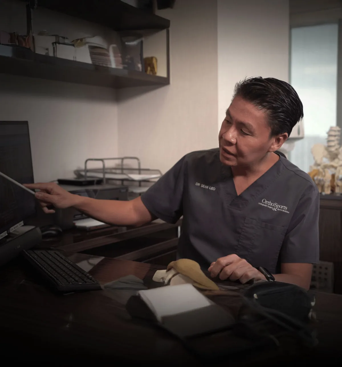
Introduction
Knee care hub · Dr Sean Leo
Orthopaedic knee care in Singapore: assessment and treatment options
Dr Sean Leo • Orthopaedic Surgeon (Specialist in Orthopaedic Surgery) • Clinical Interest: Knee Conditions, Injuries & SurgeryMBBS, MRCS (Ed), MMed (Ortho), FRCSEd (Ortho)
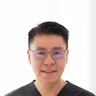
Dr Sean LeoOrthopaedic Surgeon, Singapore
Clinic enquiries · Not an emergency service
Quick linksACL injuries and reconstructionKneecap instabilityBilling and insurance
Start here
Start here
Start here
Start by understanding the knee problem, not choosing a procedure.
Knee symptoms can overlap across different conditions. This page helps you describe the concern, understand the assessment process and find relevant educational pathways without asking you to diagnose yourself.
Seeing an orthopaedic surgeon about your knee does not mean surgery has already been decided. The purpose of assessment is to understand the problem and discuss reasonable options for your situation.
OrthoKinetics is Dr Sean Leo’s professional brand. Clinical consultations and treatment are provided through OrthoSports Orthopaedic & Sports Surgery.
What’s covered on this webpage
Follow the route in order, or jump to the stop you need. Stops you have read are marked.

When should a knee problem be assessed
When should a knee problem be assessed
When should a knee problem be assessed?
Routine assessment may help when symptoms persist or limit function
A knee concern may be worth assessing when it continues, recurs or affects ordinary movement, work, recreation or sport. Describing what happened, how the knee behaves and what you can no longer do provides useful context.
Urgent concerns need an urgent-care route
- OrthoKinetics does not provide emergency or urgent care through this webpage or its WhatsApp contact.
Knee assessment
Knee assessment
What does an orthopaedic surgeon look at in a knee assessment?
Assessment brings together the history of the problem, examination of the knee and any tests that are useful for the question being considered. The exact assessment varies with the concern.
How the problem started and how the knee behaves
You may be asked when the problem began, whether there was an injury, how symptoms change and how the knee affects daily life or activity.
Movement, stability and function
Examination can consider how the knee moves, how it responds to relevant clinical tests and what the problem means for function.
Imaging when it can answer a useful question
Imaging is considered in context. A scan is one part of an assessment; it does not select treatment by itself.
A simple assessment sequence
- Describe how the problem began and how it affects you.
- Examine movement, stability and function as relevant.
- Consider targeted tests only where they may add useful information.
- Discuss the working possibilities, reasonable options and uncertainty.

Use symptoms to describe the problem, not diagnose it.
Use symptoms to describe the problem, not diagnose it.
Use symptoms to describe the problem, not diagnose it.
Pain, swelling, stiffness, catching, clicking, locking and giving way are useful descriptions. They do not confirm which structure is responsible, and more than one concern may contribute to a person’s experience.
Pain, swelling or stiffness
Describe where you feel the symptom, when it appears, what changes it and how it limits you. This gives the assessment context without turning the description into a diagnosis.
Catching, clicking or locking
Explain what you feel and whether movement is actually blocked. A sound or sensation alone should not be used to assign a condition.
Giving way or instability
Describe when the knee feels unreliable and what activity was happening at the time. Giving way does not, by itself, confirm one ligament injury.
Symptoms after a new injury
Note how the injury happened and what changed afterwards. Use the visible urgent-care guidance above if you are concerned that the problem needs prompt attention.

Knee pathways
Knee pathways
Knee pathways
Find the relevant knee pathway.
These links organise educational information by broad concern. Choosing a card does not confirm a diagnosis or mean that its treatment applies to you.

Anterior cruciate ligament (ACL) and other knee ligament injuries
Read about how ligament concerns are assessed and how rehabilitation and surgical options may be discussed. Some ligament injuries, such as ACL injuries, may follow a pathway that includes rehabilitation and, in selected situations, reconstruction using a graft.[1]

Meniscus injuries
Use the meniscus pathway for governed information about assessment and management choices. Reading it cannot confirm that a meniscus injury is causing your symptoms.
Cartilage injuries
Use the cartilage pathway for an overview of cartilage concerns and the questions that shape treatment discussions.

Kneecap instability and patellofemoral concerns
Use this pathway for information about kneecap instability and related assessment.

Tendon and other soft-tissue concerns
This route can help you find educational information about tendon or other soft-tissue concerns without treating a symptom label as a diagnosis.

Knee osteoarthritis and replacement decisions
Use this pathway for information about knee osteoarthritis and the considerations involved in partial or total knee replacement discussions. Age alone does not make replacement inevitable.

Acute trauma and combined injuries
Some injuries involve more than one structure. A pathway page can explain a topic, but individual assessment is needed to understand the whole pattern.

What tests can and cannot show
What tests can and cannot show
What tests can and cannot show
Tests can add information to a knee assessment, but no universal sequence applies to every person. Findings need to be considered alongside the history, examination and goals before treatment is discussed.
Video · Dr Sean Leo
Beyond the X-Ray: Why an MRI Can Be the Key to Your Diagnosis
- What an X-ray can and cannot show after a knee injury
- Why an MRI may be used to look at the knee's soft tissues


Treatment follows the diagnosis and the person.
Treatment follows the diagnosis and the person.
Treatment follows the diagnosis and the person.
A treatment discussion should compare reasonable non-operative and operative options where relevant. The choice depends on the knee problem, the person and the purpose of treatment; specialist review does not imply surgery.
Structure / Patient / Goal
Structure / Patient / Goal
Deciding together
Structure / Patient / Goal
S
Structure: what is affected?
Structure covers the diagnosis and the condition of the relevant joint or tissue. It includes what the assessment shows and what a test adds when testing is useful.
P
Patient: how does it affect you?
Patient means you as a whole person. Symptoms, health, function, preferences and practical circumstances can affect which options are reasonable.
G
Goal: what are you hoping to return to?
Goal describes what matters to you, such as daily independence, work, recreation or sport. A useful plan needs a clear purpose.
Your plan
Structure / Patient / Goal helps organise benefits, burdens, limitations and uncertainty without suggesting that one scan, one symptom or motivation alone can determine the outcome.

Preservation, reconstruction and replacement describe different aims.
Preservation, reconstruction and replacement describe different aims.
Preservation, reconstruction and replacement describe different aims.
These are not steps in a mandatory ladder. They describe different treatment purposes, and not every knee concern fits neatly into one category.
Preserve when appropriate
Preservation aims to retain useful tissue or joint function when that approach suits the condition, the patient and the goal.
Reconstruct when stability or tissue function needs restoration
Reconstruction aims to restore a damaged structure or its function in an appropriately selected situation. Some ligament injuries, such as ACL injuries, may involve graft reconstruction, but this is not the pathway for every ligament injury.[1]
Replace when joint damage and symptoms justify that discussion
Replacement is a separate discussion for relevant joint damage and symptoms. It is not inevitable with age, and it is not the automatic next step after another pathway.
Technology supports a plan; it does not replace one.
Technology supports a plan; it does not replace one.
Technology supports a plan; it does not replace one.
Arthroscopy is a minimally invasive surgical approach used for selected indicated knee procedures. When degeneration is too advanced for arthroscopy to be effective, this limitation will be explained.[2]
Robotic assistance is one of the technologies used in total knee replacement. Evidence comparing robotic-assisted and conventional total knee replacement shows more precise post-operative alignment with robotic techniques, while functional outcomes and complication rates are similar between the two approaches.[3]

What happens next?
What happens next?
What happens next?
Prepare for a knee assessment
Bring relevant reports or images if you have them, details of previous treatment and a current medicine list. Consider noting which activities matter most and what you want to understand.
Billing and insurance questions
Ask the clinic team for current information about fees, panel status, pre-authorisation and expected payment arrangements. Insurance coverage and out-of-pocket costs depend on the individual policy and the insurer’s decision; no coverage is guaranteed.
A cost estimate for surgery can be given only after a consultation and assessment, once a surgical plan has been developed. Each individual condition is unique — implants used, time taken, and other factors contribute to the overall estimate. The estimate will be provided to you by the clinic should you decide on surgical management, and will also be used to assist in obtaining pre-authorisation from insurance where applicable.

Insurance panels. Dr Sean Leo, Orthopaedic Surgeon, is on the specialist panels of Great Eastern (GE Panel), HSBC Life (HSBC Life Shield) and Singlife (Singlife Shield), and on the Prudential Panel. AXA Insurance Singapore became part of HSBC Life in 2023, and former AXA Shield plans are now HSBC Life Shield plans.
Corporate health plans. Dr Leo is on the panels of both Alliance Medinet and iXchange, which administer medical benefits for many company health plans. If your company plan uses either panel, you can see Dr Leo as a panel doctor.
If your company uses a different panel. Some company plans still let you see a doctor of your choice outside their panel, then claim reimbursement. This varies from plan to plan, as may the co-payment, so please check your benefits with your company’s HR department.
Panel arrangements change from time to time, so please confirm with your insurer or your company’s plan administrator before your appointment.
Dr Leo is on the panels shown. If your insurer is not listed, you may ask whether co-management with an Orthosports surgeon partner on that panel is clinically and administratively appropriate. Orthosports surgeon partners are collectively on the panels of all Shield plans.
Specific benefits, pre-authorisation, reimbursement and cashless treatment depend on the individual policy and insurer approval and are not guaranteed.
OrthoSports appointment route
Arrange a consultation: the appointment section is further down this page.

 With you through the seasons
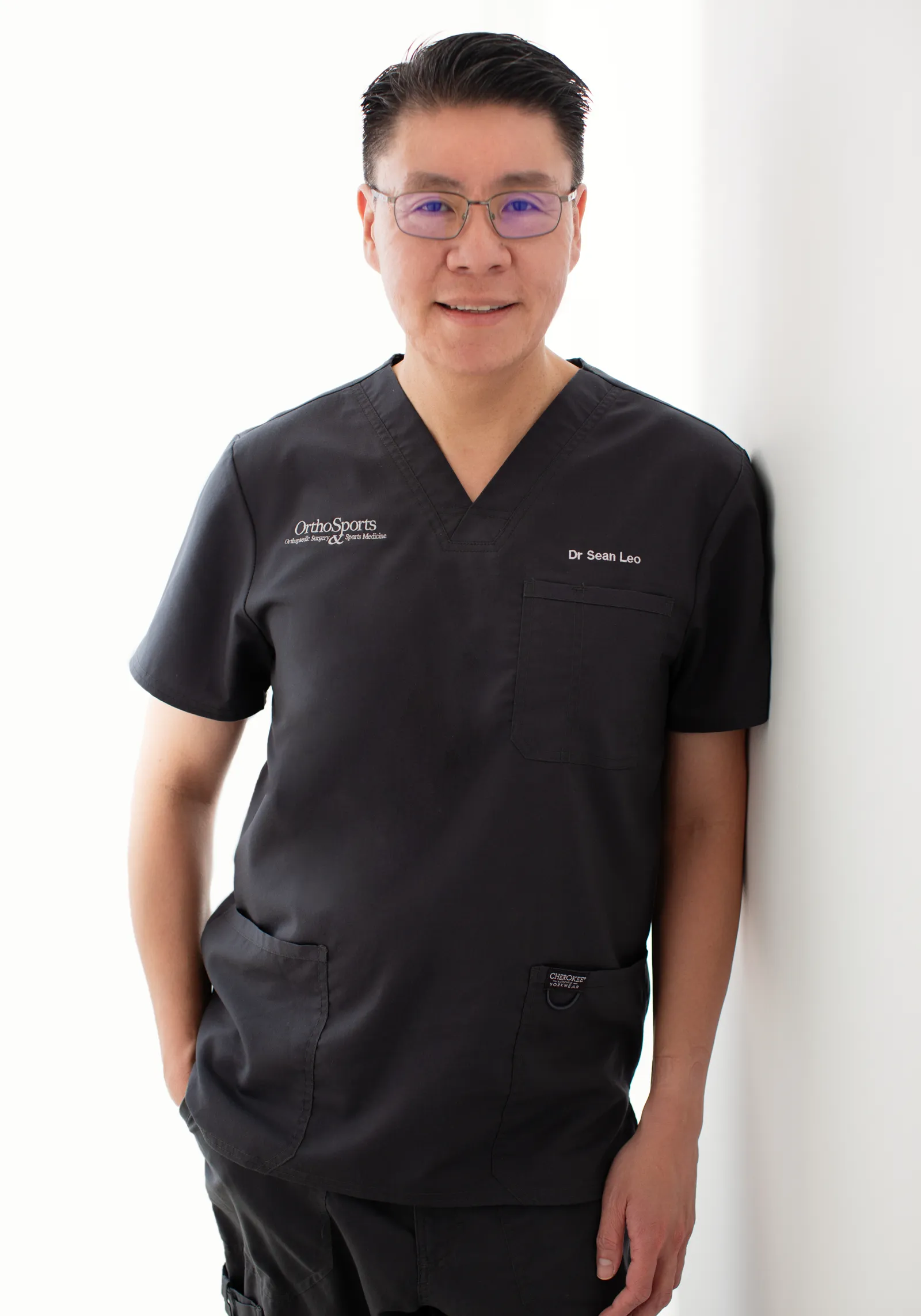
Meet Dr Sean Leo
Meet Dr Sean Leo
A consultant orthopaedic surgeon whose clinical focus is the knee, sports injuries and the lower limb.
- Dr Sean Leo is a consultant orthopaedic surgeon. OrthoKinetics is his professional brand; clinical services are provided through OrthoSports Orthopaedic & Sports Surgery at Mount Elizabeth Novena Specialist Centre and Connexion, Farrer Park Hospital.
- His clinical interests include sports injuries, including those in adolescent athletes; ligament reconstruction; realignment surgery; cartilage repair and joint-preservation approaches; knee replacement; and technology-assisted hip and knee surgery.
- The consistent aim is to return people to meaningful function and activity while avoiding unnecessary downtime.
- Qualifications: MBBS, MRCS (Ed), MMed (Ortho), FRCSEd (Ortho).
More detail: Meet Dr Sean Leo
- Training and career: Dr Leo trained in medicine at the National University of Singapore and served in the Singapore Armed Forces Medical Corps from 2000 to 2012, where his work included the care of soldiers in physically demanding roles. He completed postgraduate orthopaedic training in 2009 and a one-year clinical fellowship at Royal North Shore Hospital in Sydney, Australia, before joining Changi General Hospital as an orthopaedic consultant in 2012, where he later held the positions of Senior Consultant and Director of the Hip and Knee Service. With seniority, his educational contribution has evolved towards mentorship and support for junior doctors.

OrthoKinetics Recovery Reflection
Recovery reflection
What changes when you stop trying to name the problem yourself and begin by describing how it affects your life?
Clarity often begins with careful observation: what changed, what matters and what remains uncertain. You do not need to solve every question at once. A measured next step can begin with an honest description, then adapt as understanding grows.
Illustrative image
Questions people often ask
Common questions
Common knee questions
Do I need an MRI (magnetic resonance imaging) scan before my first knee consultation?
Not automatically. The role of imaging depends on the question being assessed. Existing reports or images may be useful, but a scan does not choose treatment by itself.
Does every ligament injury need surgery?
No. Some ligament injuries, including some ACL injuries, can be managed without surgery. Reasonable options depend on Structure / Patient / Goal and on the benefits, burdens and uncertainty relevant to the individual situation.[1]
Does every meniscus concern need arthroscopy?
No procedure should be selected from a symptom label alone. Assessment comes first. Arthroscopy has a role only for selected indications and is not effective for every degenerative knee problem.[2]
What is a truly locked knee?
A truly locked knee is one that cannot be straightened or bent through its normal range because something is mechanically blocking the joint, not simply a knee that is stiff, painful or swollen and hard to move. Whether this applies to you is a judgement for your treating clinician. If movement is blocked or you are concerned that prompt attention may be needed, use the visible urgent-care guidance above rather than relying on this page for remote triage.
Can knee symptoms involve more than one structure?
Yes. Symptom words overlap, and more than one concern may contribute. A pathway card is educational navigation, not a diagnosis.
Is knee replacement inevitable with age?
No. Preservation, reconstruction and replacement are not a mandatory ladder, and age alone does not determine treatment.
How is a treatment plan chosen?
The discussion begins with the diagnosis, the person and the purpose of treatment. Structure / Patient / Goal helps compare reasonable options, limitations and uncertainty.
See also our guide to knee pain in children and when it should be reviewed.
See also our guide to knee pain during pregnancy.
What do people mean by a “knee specialist”?
“Knee specialist” is an everyday phrase many people use when looking for a doctor for a knee problem. In Singapore, doctors may only use the specialist titles accredited by the Specialist Accreditation Board (SAB) of the Ministry of Health (MOH). Orthopaedic Surgery is an accredited specialty; the knee is an area of clinical interest within it. “Knee specialist” is not a separate specialist registration; however, individual orthopaedic surgeons may have particular clinical interests within their specialty. The doctor most people have in mind is an orthopaedic surgeon whose practice focuses on knee conditions. Dr Sean Leo is registered as a specialist in Orthopaedic Surgery, and his clinical interest is the knee. He completed a postgraduate knee fellowship programme in Sydney, Australia, at the Royal North Shore Hospital under Dr Myles Coolican and Dr David Parker from 2009 to 2010. Dr Leo was also Director of the Hip and Knee Service at Changi General Hospital from 2014 to 2018.
Video · Dr Sean Leo
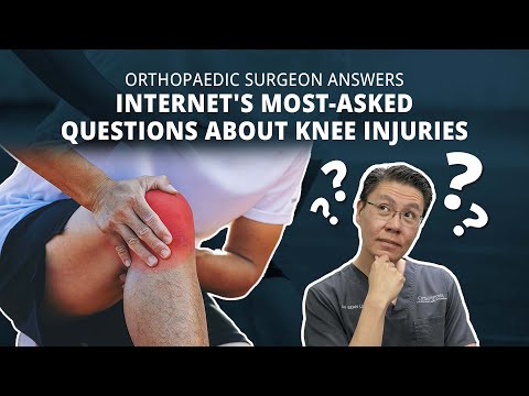
Orthopaedic Surgeon Answers Internet's MOST-ASKED Questions About KNEE PAIN (Part 1)
- How knee arthritis treatment is matched to its stage
- Why some knee injuries settle by themselves, and when assessment is sensible
Sources
References
References
- American Academy of Orthopaedic Surgeons. Management of Anterior Cruciate Ligament Injuries: Clinical Practice Guideline. Published 2022. https://www.aaos.org/globalassets/quality-and-practice-resources/anterior-cruciate-ligament-injuries/aclcpg.pdf
- Siemieniuk RAC, et al. Arthroscopic surgery for degenerative knee arthritis and meniscal tears: a clinical practice guideline. BMJ. 2017;357:j1982. doi:10.1136/bmj.j1982. https://www.bmj.com/lookup/doi/10.1136/bmj.j1982
- Alrajeb R, et al. Robotic-assisted versus conventional total knee arthroplasty: systematic review and meta-analysis of randomised controlled trials. PMID: 38133653. https://pubmed.ncbi.nlm.nih.gov/38133653/
Final next step
Final next step
Next step
Final next step
Reading about a problem is a starting point. An assessment brings together the history, an examination where needed and any useful tests, and helps you and your clinician decide what to do next. How much physical examination is needed is a matter of clinical judgement. It may be limited when pain makes an examination unreliable, or when imaging clearly shows a problem that matches the symptoms, such as a fracture seen on an X-ray or an MRI scan that clearly shows an injury consistent with the symptoms. The Singapore Medical Council and the UK General Medical Council both allow remote medical consultations, where a physical examination is not possible, with safeguards: the doctor must be clear about the limits of an opinion formed without an examination, and whether an in-person examination is needed, or referral to other services, is left to the clinical judgement of the attending doctor.
Signs that need urgent help
Some symptoms should not wait for a routine appointment. Each item below says where to go.
After an injury
- The leg looks twisted or bent, a bone is showing, there is a deep cut, or the foot or leg turns cold, pale, numb, blue or white. Go to an emergency department or call 995.
- You cannot lift your leg up with the knee kept straight, and you may feel a gap just above or just below the kneecap. Arrange an urgent same-day assessment.
At any time
- The knee is stuck and will not straighten, or it is very painful and you cannot move it or put weight on it. Arrange an urgent same-day assessment.
- The knee is suddenly very painful, hot, red and swollen, especially with a fever or feeling generally unwell. Go to an emergency department or call 995.
- One calf or thigh is newly painful and swollen. Arrange an urgent same-day assessment. If you also feel short of breath or have chest pain, go to an emergency department or call 995.
After knee surgery
- The operated knee becomes more painful, swollen or red, a wound is leaking, or you develop a fever. Go to an emergency department or call 995, or contact your surgical team straight away.
If you are admitted by the doctor at the urgent care centre of one of these private hospitals, the Parkway Pantai hospitals (Mount Elizabeth Orchard, Mount Elizabeth Novena, Gleneagles and Parkway East), Farrer Park Hospital and Thomson Medical Centre, you may ask for Dr Sean Leo to be your attending orthopaedic surgeon. If you are instead discharged and referred for orthopaedic follow-up, you may contact the clinic to arrange an appointment. Both are subject to the hospital’s admission processes and to Dr Leo’s availability.
Arrange a consultation
Appointments are arranged through the OrthoSports clinic team. Send your details below, or message or call the clinic.
Leave your details and the clinic team will reply to arrange a time.
Message the clinic on WhatsApp
-
38 Irrawaddy Road, #10-41 Mount Elizabeth Novena Specialist Centre, Singapore 329563 Map for Orthosports@Novena (opens Google Maps)
-
1 Farrer Park Station Road, #15-17 Connexion, Singapore 217562 Map for Orthosports@Connexion (opens Google Maps)
WhatsApp is for clinic enquiries only. It is not an emergency service and is not monitored for urgent care. Website or messaging contact is not an emergency-response service. For a medical emergency, use your local emergency service.
- OrthoKinetics does not provide emergency or urgent care through this webpage or its WhatsApp contact.