
Introduction
Sports injuries · Jumper’s knee
Patellar tendonitis (jumper’s knee): front-of-knee pain, assessment and treatment
Written by Dr Sean Leo, a consultant orthopaedic surgeon whose clinical focus is the knee, sports injuries and the lower limb.
Last updated: 9 October 2026.

Dr Sean LeoOrthopaedic Surgeon, Singapore
Clinic enquiries · Not an emergency service
Illustrative image

What is patellar tendonitis
What is patellar tendonitis
What is patellar tendonitis (jumper’s knee)?
- Patellar tendonitis or jumper’s knee (patellar tendinopathy) is pain in the tendon just below the kneecap. It usually builds up gradually with repeated jumping, landing and running, rather than after a single injury.[1][3]
- The first plan is usually to adjust the load and build the tendon back up gradually. It is not complete rest, and surgery is the exception.[2][3]
- A sudden, severe injury that stops you straightening the knee is a different, acute problem and should be assessed promptly.[4][6]
- OrthoKinetics is Dr Sean Leo’s professional brand. Clinical consultations and treatment are provided through OrthoSports Orthopaedic & Sports Surgery. Dr Sean Leo is a specialist orthopaedic surgeon who cares for sports injuries.
What’s covered on this webpage
Follow the route in order, or jump to the stop you need. Stops you have read are marked.
What is the patellar tendon, and where does it attach
What is the patellar tendon, and where does it attach
What is the patellar tendon, and where does it attach?
The patellar tendon attaches the bottom of the kneecap (the patella) to the top of the shinbone (the tibia).[4] It is the tendon below the kneecap, and it is the one this page is about.
The quadriceps tendon is a different structure. It connects the thigh muscle to the top of the kneecap.[5] So the location of the pain is a useful clue: pain in the tendon below the kneecap points to the patellar tendon, and pain in the tendon above it points to another tendon. If your pain sits above the kneecap, the page on pain above the kneecap (quadriceps tendonitis) may be the more relevant one to read.[5]
Why is it sometimes called a ligament?
A structure that connects one bone to another is technically a ligament, so the patellar tendon is sometimes called the patellar ligament.[4]

What brings on patellar tendonitis
What brings on patellar tendonitis
What brings on patellar tendonitis?
Jumper’s knee is a painful, mainly activity-related problem of the patellar tendon. It comes from stress that builds up in the tendon over time, and it is not usually the result of one injury.[1]
What is commonly called “tendonitis” is better understood as a gradual, load-driven tendon problem, known medically as tendinopathy, than as a simple, short-lived inflammation.[2] Tendon problems of this kind sit on a continuum, and adding or removing load is the main thing that moves the tendon forward or back along it.[2] That is why treatment focuses on load: the tendon is reacting to how it has been used, and that is something that can change.[2][3]
A sudden rise in jumping and landing can be enough to bring it on. A young jumping athlete who greatly increases the number of jumps and landings each week may develop swelling and pain in the tendon.[2]
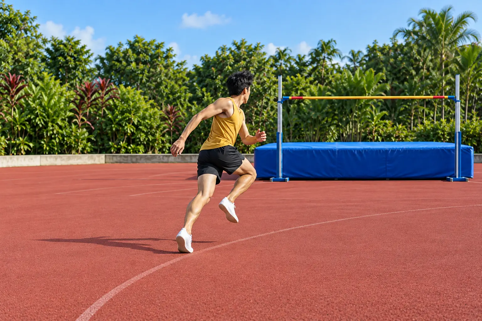
The people most often affected are those who jump, land and run a lot, in any sport. It is common in volleyball, basketball, long jump and high jump, distance running and skiing.[1] The list is only a guide. It affects recreational athletes as well as professionals.[3]
It is most common in men, with a higher rate among teenagers and young adults.[1]
Several risk factors have been described:
- Taking part in jumping sports and training on hard surfaces are listed as risk factors.[7]
- Greater activity volume, higher body weight, less ankle movement (dorsiflexion) and less flexibility in the muscles at the back and front of the thigh are listed as factors that still need more evidence.[7]
- Recent use of fluoroquinolone antibiotics or an underlying connective tissue disease are also named as possible risk factors.[7] Check with your doctor or pharmacist before taking any medicine.
How common is it?
Studies of particular groups of athletes give a sense of the scale. In one study, about 45% of professional volleyball players and 32% of professional basketball players were affected. In a large group of non-elite athletes across seven sports, about 8.5% were affected overall, with the highest figure in volleyball players at 14.4%.[3] These are figures for specific groups of athletes. They are not a measure of your own risk.
What are the symptoms of patellar tendonitis
What are the symptoms of patellar tendonitis
What are the symptoms of patellar tendonitis?
Most athletes with this problem describe pain at one clear spot at the front of the knee, at the patellar tendon.[3] People commonly describe a pattern like this:[3]
- the pain is often worse with exercise, and occasionally after the knee has been kept bent for a long time, such as when sitting;
- it often starts gradually, and people often link it to a period of increased sport;
- in mild cases it appears only during sport;
- as the problem progresses, it is often felt at the beginning of exercise or throughout it;
- in more severe cases it is felt during daily activities, or even at rest.
These are useful clues for an assessment. Front-of-knee pain on its own does not confirm which structure is responsible, because other problems at the front of the knee can feel similar.[3]
Is it jumper’s knee, or a different problem at the front of the knee
Is it jumper’s knee, or a different problem at the front of the knee
Is it jumper’s knee, or a different problem at the front of the knee?
Three different problems can be mistaken for one another. They lead in different directions.
- Pain above the kneecap. This points to the quadriceps tendon, a different tendon from the patellar tendon.[5] It has its own page: quadriceps tendonitis, pain above the kneecap.
- A kneecap that slips or gives way. This is kneecap instability, a different problem from tendon overload. It is covered on the MPFL reconstruction surgery and kneecap realignment surgery pages.
- A sudden tear of the patellar tendon. This is an injury during activity, with sudden, severe pain, and it is not gradual overuse. A completely torn patellar tendon is separated from the kneecap, and without that attachment you cannot straighten your knee.[4][6] It takes a very strong force to tear it.[4] A sudden injury like this is a reason for prompt assessment. The signs that need urgent help are listed in the Final next step section at the end of this page.
This page is not a way to check yourself from a distance. An assessment is how any uncertainty is settled. Other sports injuries are gathered on the sports injury hub.

When should I get this assessed?
When should I get this assessed?
When should I get this assessed?
Many people try managing a sore tendon themselves first, and that is a reasonable place to start. Knee symptoms after an injury that do not improve within a few weeks are a good reason to see a clinician.[6] So is being unsure what the problem is.
A different situation needs a quicker response. If the pain is sudden and severe and comes on during an accident or activity, you may have torn a tendon.[6] If you also cannot straighten your knee, this may be a torn patellar tendon and it should be assessed promptly.[4]
Safety note: This webpage is not an emergency service. A sudden, severe injury that stops you straightening your knee should not wait. See the signs that need urgent help in the Final next step section. No list on this page can rule out a serious problem remotely.
How is patellar tendonitis assessed
How is patellar tendonitis assessed
How is patellar tendonitis assessed?
The diagnosis is made mainly from the clinical picture.[3] It starts with your story: pain that came on gradually and is tied to load. Then comes an examination. The clinician looks for tenderness at the patellar tendon, typically at the lower end of the kneecap, and for how well you tolerate loaded activity.[1][3]
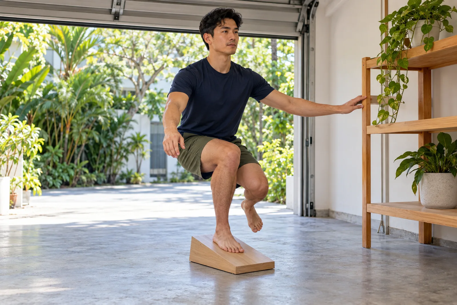
One example is the single-leg decline squat. In it you squat on one leg on a surface that slopes downward. People with this problem can often manage only a limited number before the pain starts, which suggests the tendon is sensitive to load.[3] It is an example of a loaded test used in an examination. It is not a test to use on yourself at home.
A scan can support the assessment, but it does not replace the story and the examination.[3]
What scans add
Ultrasound and MRI are the most useful scans for looking at a tendon, because they show it in detail. An ultrasound can show an area of tendon thickening.[3] An X-ray shows bone.[3] Scan changes can also be present in athletes who have no symptoms, so a scan finding is not the diagnosis on its own. What a scan shows does not by itself decide the treatment.[3]
What is the main treatment for patellar tendonitis
What is the main treatment for patellar tendonitis
What is the main treatment for patellar tendonitis?
Treatment without surgery is the first choice, and its mainstay is progressive tendon-loading exercise.[3] Eccentric exercise, in which the leg is lowered under load, is the form studied most widely.[3] A recent systematic review found that combining load management with exercise can give favourable results in both the short and long term.[3]
A 2026 UK Defence Rehabilitation consensus panel, working with a military population, strongly advocates exercise rehabilitation as the primary management of Achilles and patellar tendinopathy.[8]
Load is what drives the problem, so it is also part of the answer. The aim is to adjust your training load and volume, not to stop everything and rest for a long stretch.[2][3] How much to adjust is a clinical judgement worked out with your clinician. No single exercise or protocol suits everyone, and other loading approaches are also used.[3]
Other measures play smaller roles:
- Anti-inflammatory medicines have a limited and debated role. They are not the main treatment.[3] If you take a prescribed medicine, do not stop it without speaking to your clinician. Non-steroidal anti-inflammatory drugs (NSAIDs) are not associated with long-term improvement in clinical outcomes or tendon structure, but may offer some short-term pain relief.[8] The side-effect profile of these medicines must be considered.[8] NSAIDs and acetaminophen (paracetamol) may be used sparingly for short-term pain management.[7] Check with your doctor or pharmacist before taking any medicine.
- Steroid (cortisone) injections may give short-term relief at best. They are not better than other treatments in the long term, and they have been linked to a weakened tendon and a greater likelihood of the tendon rupturing. Doctors typically avoid giving them in or around the patellar tendon.[3][4]
- Injecting steroid into the tendon has been associated with a risk of tendon rupture.[8] The consensus graded this as Level 4 evidence.[8] On the Oxford evidence scale it uses, Level 4 means case series, case-control or historically controlled studies, so this link has not been robustly proven in studies.[9]
When is surgery considered?
When is surgery considered?
When is surgery considered?
Many athletes respond well to treatment without surgery.[3] Surgery is considered when someone has not improved after a sustained period of that treatment, commonly cited as about 6 months.[3] There is no clear evidence about which surgical approach works best.[3]
Structure / Patient / Goal
Structure / Patient / Goal
Deciding together
Structure / Patient / Goal
S
Structure: what is affected?
Structure is the patellar tendon itself: where it sits below the kneecap, what the assessment shows, and how the rest of the knee around it is involved. It also includes what a scan adds, when a scan is useful.
P
Patient: how does it affect you?
Patient means you as a whole athlete and person. Your symptoms, how active you are, your general health, your preferences and your circumstances all shape which options are reasonable.
G
Goal: what are you hoping to return to?
Goal is what matters to you, whether that is daily activity, work, running or a jumping sport. A useful plan starts from a clear purpose.
Your plan
No single factor decides treatment alone, and one scan or one symptom does not determine the plan.
What does surgery for patellar tendonitis involve
What does surgery for patellar tendonitis involve
What does surgery for patellar tendonitis involve?
The operation removes damaged tendon tissue.[6] It can be done through an open incision or arthroscopically, which is the keyhole approach. Arthroscopic techniques are more commonly preferred because they are less invasive.[3]
There is no clear evidence that any one technique is better than another, and surgery does not guarantee a pain-free result.[3]
What does recovery from patellar tendonitis involve
What does recovery from patellar tendonitis involve
What does recovery from patellar tendonitis involve?
Tendons respond slowly, both in gaining load capacity and in settling pain.[2] Because a tendon can move up and down between stages, estimating how long recovery will take is difficult.[2] For that reason progress is judged by how the tendon copes as the load increases, not by a date on the calendar.[2]
Many people respond well to non-operative treatment.[3] The condition can also persist for years, so expectations are set honestly from the start.[3] Return to sport happens in steps, guided by how the tendon responds. It does not follow a fixed date, and it is not a promise of returning to your previous level.[2][3]
Risks and complications
Risks and complications
Risks and complications
Both paths carry real limits.
Non-operative care. Anti-inflammatory medicines have a limited and debated role, and some evidence suggests they may impair tendon healing.[3] Check with your doctor or pharmacist before taking any medicine. Steroid injections give short-term relief at best and have been linked to tendon weakness and a greater likelihood of the tendon rupturing, so they are typically avoided in or around the patellar tendon.[3][4] The condition can persist even with treatment.[3]
Surgery. There is no clear evidence for one best surgical approach, and no result is guaranteed.[3] Most people improve, but not everyone does.[3]

OrthoKinetics Recovery Reflection
Recovery reflection
If the tendon below your kneecap is the spring that powers your jump, what does it mean to re-learn to trust it one gentler landing at a time?
It can help to picture the tendon as a spring: it takes in the force of every landing and gives it back in the next jump. A spring that has been overworked is not asked to bounce as high as ever on its first day back. It is asked for gentler landings, practised often. Re-learning to trust it can feel less like waiting to heal and more like rehearsal.
What changes when a lighter session counts as progress in its own right, rather than as a sign that you can return to full load straight away?
Athletes are often taught to read a good day as a green light. A tendon that is being rebuilt keeps a different kind of score. A lighter session that goes well has done its job by being lighter. Seeing it that way can take some of the frustration out of the days that feel like holding back, because those days are part of how the spring is rebuilt.
A spring works by giving a little and returning a little, over and over. A tendon being rebuilt is asked to learn the same rhythm, one landing after another.
Illustrative image

 With you through the seasons
Meet Dr Sean Leo
Meet Dr Sean Leo
A consultant orthopaedic surgeon whose clinical focus is the knee, sports injuries and the lower limb.
- Dr Sean Leo is a consultant orthopaedic surgeon. OrthoKinetics is his professional brand; clinical services are provided through OrthoSports Orthopaedic & Sports Surgery at Mount Elizabeth Novena Specialist Centre and Connexion, Farrer Park Hospital.
- His clinical interests include sports injuries, including those in adolescent athletes; ligament reconstruction; realignment surgery; cartilage repair and joint-preservation approaches; knee replacement; and technology-assisted hip and knee surgery.
- The consistent aim is to return people to meaningful function and activity while avoiding unnecessary downtime.
- Qualifications: MBBS, MRCS (Ed), MMed (Ortho), FRCSEd (Ortho).
More detail: Meet Dr Sean Leo
- Training and career: Dr Leo trained in medicine at the National University of Singapore and served in the Singapore Armed Forces Medical Corps from 2000 to 2012, where his work included the care of soldiers in physically demanding roles. He completed postgraduate orthopaedic training in 2009 and a one-year clinical fellowship at Royal North Shore Hospital in Sydney, Australia, before joining Changi General Hospital as an orthopaedic consultant in 2012, where he later held the positions of Senior Consultant and Director of the Hip and Knee Service. With seniority, his educational contribution has evolved towards mentorship and support for junior doctors.
Questions people often ask
Common jumper’s knee questions
Common jumper’s knee questions
Is jumper’s knee the same as patellar tendonitis?
Yes. Jumper’s knee, patellar tendonitis and patellar tendinopathy all describe a painful, mainly activity-related problem of the tendon below the kneecap. “Tendinopathy” is the more accurate medical term, because it is usually a gradual, load-driven tendon problem rather than a simple, short-lived inflammation.[1][2]
What causes pain just below the kneecap?
Pain just below the kneecap often comes from the patellar tendon, which joins the kneecap to the shinbone. It typically appears at one clear spot and is worse with exercise. Other problems at the front of the knee can feel similar, so an assessment is what confirms the cause.[1][3][4]
What causes jumper’s knee?
It comes from stress that builds up in the tendon over time, rather than from one injury. A sudden rise in jumping and landing can bring it on. It is common in jumping and running sports, such as volleyball, basketball and distance running, and it affects recreational athletes too.[1][2][3]
Do I need to stop my sport completely?
Usually the aim is to adjust your load and training volume, not to stop everything. Load is what drives the problem, so managing it is part of how the tendon settles. How much to adjust is a clinical judgement worked out with your clinician.[2][3]
What exercises help patellar tendonitis?
Progressive tendon-loading exercise is the mainstay of treatment. Eccentric exercise, in which the leg is lowered under load, is the form studied most widely, and other loading approaches are also used. Your clinician chooses the programme, because no single exercise or protocol suits everyone.[3]
Should I rest a sore patellar tendon completely?
Complete, prolonged rest is not the usual approach. The tendon responds to load, so treatment adjusts your training load and volume and then builds the tendon’s tolerance back up gradually. How much activity suits you at each stage is worked out with your clinician.[2][3]
Do I need an MRI for patellar tendonitis?
Not to make the diagnosis. The diagnosis is made mainly from your story and an examination. Ultrasound and MRI can show detail in the tendon and support the assessment, but they do not replace it, and a scan finding does not by itself decide treatment.[3]
What is the difference between patellar and quadriceps tendonitis?
They affect different tendons. Patellar tendonitis affects the tendon below the kneecap, which joins it to the shinbone. Quadriceps tendonitis affects the tendon above the kneecap, which connects the thigh muscle to it. There is a separate page on quadriceps tendonitis.[4][5]
Will I need surgery for jumper’s knee?
Surgery is the exception. Many people respond well to non-operative treatment. Surgery is considered only when someone has not improved after a sustained period of that treatment, commonly cited as about 6 months, and there is no clear evidence for one best surgical approach.[3]
How long does jumper’s knee take to heal?
There is no fixed timeline. Tendons respond slowly, so recovery is gradual and varies from person to person, and progress is guided by how the tendon copes as the load increases. The condition can also persist for years, so expectations are set honestly.[2][3]
What do people mean by a “knee specialist”?
“Knee specialist” is an everyday phrase many people use when looking for a doctor for a knee problem. In Singapore, doctors may only use the specialist titles accredited by the Specialist Accreditation Board (SAB) of the Ministry of Health (MOH). Orthopaedic Surgery is an accredited specialty; the knee is an area of clinical interest within it. “Knee specialist” is not a separate specialist registration; however, individual orthopaedic surgeons may have particular clinical interests within their specialty. The doctor most people have in mind is an orthopaedic surgeon whose practice focuses on knee conditions. Dr Sean Leo is registered as a specialist in Orthopaedic Surgery, and his clinical interest is the knee. He completed a postgraduate knee fellowship programme in Sydney, Australia, at the Royal North Shore Hospital under Dr Myles Coolican and Dr David Parker from 2009 to 2010. Dr Leo was also Director of the Hip and Knee Service at Changi General Hospital from 2014 to 2018.
Costs and insurance
Costs and insurance
Practicalities
Cost, MediSave and insurance

Ask the clinic team for current information about fees, panel status, pre-authorisation and expected payment arrangements. Insurance coverage and out-of-pocket costs depend on the individual policy and the insurer’s decision; no coverage is guaranteed.
A cost estimate for surgery can be given only after a consultation and assessment, once a surgical plan has been developed. Each individual condition is unique — implants used, time taken, and other factors contribute to the overall estimate. The estimate will be provided to you by the clinic should you decide on surgical management, and will also be used to assist in obtaining pre-authorisation from insurance where applicable.
Insurance panels. Dr Sean Leo, Orthopaedic Surgeon, is on the specialist panels of Great Eastern (GE Panel), HSBC Life (HSBC Life Shield) and Singlife (Singlife Shield), and on the Prudential Panel. AXA Insurance Singapore became part of HSBC Life in 2023, and former AXA Shield plans are now HSBC Life Shield plans.
Corporate health plans. Dr Leo is on the panels of both Alliance Medinet and iXchange, which administer medical benefits for many company health plans. If your company plan uses either panel, you can see Dr Leo as a panel doctor.
If your company uses a different panel. Some company plans still let you see a doctor of your choice outside their panel, then claim reimbursement. This varies from plan to plan, as may the co-payment, so please check your benefits with your company’s HR department.
Panel arrangements change from time to time, so please confirm with your insurer or your company’s plan administrator before your appointment.
Dr Leo is on the panels shown. If your insurer is not listed, you may ask whether co-management with an Orthosports surgeon partner on that panel is clinically and administratively appropriate. Orthosports surgeon partners are collectively on the panels of all Shield plans.
Specific benefits, pre-authorisation, reimbursement and cashless treatment depend on the individual policy and insurer approval and are not guaranteed.
Arrange a consultation: the appointment section is further down this page.
Sources
References
References
- Mabrouk A, Lee CYM, Sherman AL. Patellar Tendinopathy (Jumper’s Knee). [2026 Jan]. In: StatPearls [Internet]. Treasure Island (FL): StatPearls Publishing. NBK532969. PMID 30422564. Available at: https://www.ncbi.nlm.nih.gov/books/NBK532969/
- Cook JL, Purdam CR. Is tendon pathology a continuum? A pathology model to explain the clinical presentation of load-induced tendinopathy. Br J Sports Med. 2009;43(6):409-16. doi:10.1136/bjsm.2008.051193. PMID 18812414.
- Theodorou A, Komnos G, Hantes M. Patellar tendinopathy: an overview of prevalence, risk factors, screening, diagnosis, treatment and prevention. Arch Orthop Trauma Surg. 2023;143(11):6695-6705. doi:10.1007/s00402-023-04998-5. PMID 37542006. PMCID PMC10541843.
- American Academy of Orthopaedic Surgeons (AAOS). Patellar Tendon Tear. OrthoInfo. Available at: https://orthoinfo.aaos.org/en/diseases–conditions/patellar-tendon-tear
- American Academy of Orthopaedic Surgeons (AAOS). Quadriceps Tendon Tear. OrthoInfo. Available at: https://orthoinfo.aaos.org/en/diseases–conditions/quadriceps-tendon-tear
- NHS. Tendonitis. Available at: https://www.nhs.uk/conditions/tendonitis/
- Walton J, Kozina E, et al. A Review of Patellar Tendinopathy in Athletes Involved in Jumping Sports. Cureus. 2023;15:e47459. doi:10.7759/cureus.47459. PMID:38022235.
- Judd, Wild, et al. UK Defence Rehabilitation consensus agreement for the conservative management of Achilles and patellar tendinopathy. BMJ Mil Health. 2026;172(2):172-177. doi:10.1136/military-2024-002893. PMID:39824541.
- Howick J, Glasziou P, Greenhalgh T, et al. The Oxford 2011 Levels of Evidence. Oxford Centre for Evidence-Based Medicine. https://www.cebm.ox.ac.uk/resources/levels-of-evidence/ocebm-levels-of-evidence
Final next step
Final next step
Next step
Final next step
Reading about a problem is a starting point. An assessment brings together the history, an examination where needed and any useful tests, and helps you and your clinician decide what to do next. How much physical examination is needed is a matter of clinical judgement. It may be limited when pain makes an examination unreliable, or when imaging clearly shows a problem that matches the symptoms, such as a fracture seen on an X-ray or an MRI scan that clearly shows an injury consistent with the symptoms. The Singapore Medical Council and the UK General Medical Council both allow remote medical consultations, where a physical examination is not possible, with safeguards: the doctor must be clear about the limits of an opinion formed without an examination, and whether an in-person examination is needed, or referral to other services, is left to the clinical judgement of the attending doctor.
Signs that need urgent help
Some symptoms should not wait for a routine appointment. Each item below says where to go.
After an injury
- The kneecap has slipped out of place and is still out, or the knee looks deformed. Go to an emergency department. If the kneecap went back into place by itself, arrange an early assessment.
- The leg looks twisted or bent, a bone is showing, there is a deep cut, or the foot or leg turns cold, pale, numb, blue or white. Go to an emergency department or call 995.
- You cannot lift your leg up with the knee kept straight, and you may feel a gap just above or just below the kneecap. Arrange an urgent same-day assessment.
At any time
- The knee is stuck and will not straighten, or it is very painful and you cannot move it or put weight on it. Arrange an urgent same-day assessment.
- The knee is suddenly very painful, hot, red and swollen, especially with a fever or feeling generally unwell. Go to an emergency department or call 995.
- One calf or thigh is newly painful and swollen. Arrange an urgent same-day assessment. If you also feel short of breath or have chest pain, call 995.
After knee surgery
- The operated knee becomes more painful, swollen or red, a wound is leaking, or you develop a fever. Contact your surgical team straight away. If you cannot reach them the same day, go to an emergency department. If you feel very unwell, go to an emergency department. If you are short of breath or have chest pain, call 995.
If you are admitted by the doctor at the urgent care centre of one of these private hospitals, the Parkway Pantai hospitals (Mount Elizabeth Orchard, Mount Elizabeth Novena, Gleneagles and Parkway East), Farrer Park Hospital and Thomson Medical Centre, you may ask for Dr Sean Leo to be your attending orthopaedic surgeon. If you are instead discharged and referred for orthopaedic follow-up, you may contact the clinic to arrange an appointment. Both are subject to the hospital’s admission processes and to Dr Leo’s availability.
Was this page helpful?
Arrange a consultation
Appointments are arranged through the OrthoSports clinic team. Send your details below, or message or call the clinic.
Leave your details and the clinic team will reply to arrange a time.
Message the clinic on WhatsApp
-
38 Irrawaddy Road, #10-41 Mount Elizabeth Novena Specialist Centre, Singapore 329563 Map for Orthosports@Novena (opens Google Maps)
-
1 Farrer Park Station Road, #15-17 Connexion, Singapore 217562 Map for Orthosports@Connexion (opens Google Maps)
WhatsApp is for clinic enquiries only. It is not an emergency service and is not monitored for urgent care. Website or messaging contact is not an emergency-response service. For a medical emergency, use your local emergency service.
- OrthoKinetics does not provide emergency or urgent care through this webpage or its WhatsApp contact.