appointment@orthokinetics.sg
Emergency Call +65 8439 2120

That grinding sensation with every step. The persistent ache limiting your movement. The instability makes you hesitate before pivoting. When knee cartilage wears away, every activity becomes a challenge.
Dr Sean Leo draws on 25+ years of clinical experience with subspecialty expertise in knee and lower limb surgery to restore smooth, confident knee function through advanced cartilage repair techniques proven effective for Singapore’s active adults.
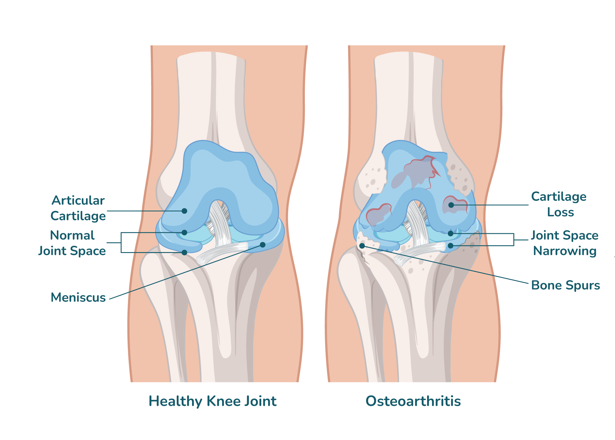
Picture the smooth, glistening tissue allowing your knee to bend and bear weight without friction—that’s articular cartilage. When this protective layer deteriorates through injury or years of activity, bone grinds against bone, triggering inflammation and progressive damage.
Unlike bone, knee cartilage lacks blood supply so damaged cartilage cannot heal naturally. This biological limitation makes early intervention essential for preventing progressive joint destruction.
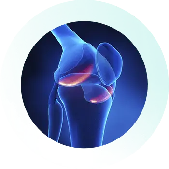
Deep, aching pain felt along the joint line, triggered by squatting, twisting, or weight-bearing activities. Swelling develops within 24-48 hours and may persist, indicating ongoing inflammation from damaged cartilage surfaces.
The knee becomes tender when touched, especially along the joint line. This deep, localised discomfort differs from muscle soreness as it persists even after rest periods and indicates cartilage breakdown.
Difficulty fully bending or straightening the knee interferes with kneeling, climbing stairs, or standing from chairs. Morning stiffness lasting 30+ minutes reflects inflammation and irregular joint surfaces from cartilage wear.
Knee cartilage wear or loose fragments disrupt smooth joint motion, causing catching, clicking, or locking during movement. True locking, where the knee gets stuck, requires immediate specialist evaluation.
Distinctive grinding, crunching, or popping felt during movement as rough cartilage surfaces rub together. Crepitus accompanied by pain indicates significant cartilage loss warranting assessment.
The severity of symptoms doesn’t always correlate with damage extent. Small knee cartilage wear in critical locations can produce significant pain.
Seek immediate assessment: True locking, severe rapid swelling, inability to bear weight
Schedule evaluation within 1-2 weeks: Persistent pain limiting activities, grinding with movement, progressive stiffness
Consider specialist consultation: Symptoms beyond 2-3 weeks, interference with work/activities, previous knee injuries
Early cartilage repair intervention prevents progressive destruction. Manageable discomfort can escalate to debilitating pain if knee cartilage wear continues unchecked.
Cartilage injury treatment depends on the severity and location of the damage. Mild cases may be managed with rest, physiotherapy, and activity modification. For more serious injuries, advanced options like cartilage repair, regeneration procedures, or minimally invasive surgery may be recommended to restore joint function and prevent long-term damage.
Cartilage repair surgery may be necessary for individuals with significant cartilage damage that does not improve with conservative treatments. Factors that suggest the need for surgery include:
Early evaluation by an orthopedic specialist is essential to determine if surgery is the best option based on the type and severity of the injury.
Recovery from cartilage repair surgery typically takes 3 to 6 months, depending on the severity of the injury, the surgical technique used, and individual healing. A structured rehabilitation program is crucial for optimal recovery.
The recovery process generally follows these phases:
Initial phase (0–2 weeks)
Focuses on protecting the repair site, managing swelling, and starting gentle movement with crutches or a brace.
Early rehabilitation (2–6 weeks)
Involves increasing range of motion and starting light strengthening exercises under medical supervision.
Intermediate phase (6–12 weeks)
Aims to restore strength, balance, and functional movement for daily activities.
Return to activity (3–6 months)
Patients may resume low-impact sports and other activities, guided by their orthopedic specialist.
Regular follow-ups and adherence to the rehabilitation plan are essential for the best results.
When cartilage repair techniques match your defect’s characteristics, most people experience meaningful improvement. Studies tracking patients over several years show significant pain reduction (from severe 7/10 discomfort to mild 2/10) and substantial functional gains, with quality of life scores improving from 50 to 85 out of 100.
This recent systematic review analysed multiple cartilage repair techniques, including ACI, MACI, osteochondral autograft, and allograft and pooled clinical outcomes across studies. The findings show that these biological repair methods improved knee function and reduced pain, especially when defect characteristics and patient factors were appropriately considered. This reinforces that cartilage repair is not just structurally plausible but effectively felt by patients during daily movements.
Title: Autologous chondrocyte implantation, matrix-induced autologous chondrocyte implantation, osteochondral autograft transplantation and osteochondral allograft improve knee function and pain with considerations for patient and cartilage defect characteristics: A systematic review and meta-analysis
Authors: Joseph E. Nassar, Grace Guerin, Taidhgin Keel, Raffaella Russo, Filippo Familiari, et al.
Published in: Knee Surgery, Sports Traumatology, Arthroscopy
Publication Year: 2024
This systematic review shows that cartilage repair procedures can produce consistent improvement in clinical outcomes like pain and function, supporting real-world effectiveness for patients seeking cartilage repair and options.
Read the full cartilage repair outcomes review here →
PMCID: PMC12310097
In a case of a series of patients undergoing autologous chondrocyte patch implantation for knee cartilage defects, researchers observed postoperative improvements in both clinical scores and imaging measures. Patients reported decreased pain and better knee function at follow-up, and MRI evaluations showed more continuous cartilage repair tissue, meaning the physical structure of the joint was visibly improving.
Title: Four cases report: Treatment of knee joint cartilage defects using autologous chondrocyte patch implantation
Authors: Le Wang, Han Li, Yiguo Cao, Cheng Song, Qi Chen, Jun Hao, Weiguo Zhang, Kang Tian
Published in: Frontiers in Surgery
Publication Year: 2022
This case series demonstrates that biologic cartilage repair techniques can lead to observable improvements in joint cartilage and patient-reported outcomes, giving a visual and clinical sense of restored cartilage in the knee.
Review the clinical cartilage repair case series here →
PMCID: PMC9679023
This systematic review focused on the effectiveness and safety of autologous chondrocyte implantation in younger patients with cartilage defects. It found that ACI can be effective in improving knee function and reducing pain in skeletally immature individuals, extending the evidence base beyond adult populations.
Title: Autologous chondrocyte implantation in the knee is effective in skeletally immature patients: a systematic review
Authors: Filippo Migliorini, Joerg Eschweiler, Julia Prinz, Christian David Weber, Ulf Krister Hofmann, Frank Hildebrand, Nicola Maffulli
Published in: Knee Surgery, Sports Traumatology, Arthroscopy
Publication Year: 2022
This study supports the broader effectiveness of cartilage repair techniques and suggests results can be seen across age groups, including those with developing cartilage. Echoing the functional improvements felt during movement and daily activities after successful repair.
Explore the full systematic review on cartilage repair in immature knees here →
PMCID: PMC10183428
These PMCID-accessible research articles show that:
This evidence supports the claim that when cartilage repair is performed appropriately — considering defect characteristics, patient age, and clinical context — patients can move more comfortably and experience sustained improvements, which is especially relevant for people exploring treatments like cartilage repair Singapore.
The cost of cartilage repair surgery varies depending on the complexity of the procedure and individual insurance coverage. Dr. Sean’s clinic staff will discuss the specific cost details with you after your consultation if you choose to proceed with surgery. They are experienced in assisting with applications for letters of guarantee from various insurance panels and corporate insurance plans.
Factors influencing the cost include:
The clinic will provide a clear breakdown of the costs based on your treatment plan and insurance coverage.
Dr Sean Leo’s subspecialty expertise in knee and lower limb surgery ensures your knee cartilage receives specialised attention.
Expertise across multiple cartilage restoration approaches from AMIC to osteochondral grafting, enables personalised technique selection based on your specific defect characteristics.
Recommendations grounded in clinical research, ensuring approaches with demonstrated efficacy rather than experimental techniques.
Close physiotherapy collaboration ensures rehabilitation protocols match your surgical technique, optimising biological healing.
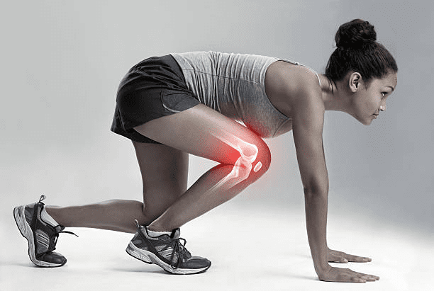
Here’s the honest truth: your knee cartilage can’t truly regenerate on its own because it lacks the blood supply needed to deliver healing cells and nutrients. But there’s real hope. Modern cartilage repair techniques like AMIC help your body create new cartilage-like tissue that restores the smooth gliding you’ve been missing. Think of it as guiding your body’s own healing potential: we stimulate bone marrow cells to migrate into the damaged area, then support them with a protective scaffold that helps new tissue form and mature. Whilst this new tissue isn’t identical to your original cartilage, it cushions your joint beautifully, reduces pain, and lets you move confidently again. The key is addressing knee cartilage damage early, before bone-on-bone wear develops. Giving your knee the best chance to heal whilst joint-preserving options still work.
You’ll recognise knee cartilage damage through that deep, persistent aching that intensifies when you’re walking, climbing stairs, or simply standing. It’s different from a sharp, sudden pain. Many people notice grinding or crunching sensations with movement, swelling that appears after activity, and stiffness that makes getting up after rest uncomfortable. Some experience catching or locking if cartilage fragments break loose, that unsettling instability during direction changes, or reduced motion that makes squatting and kneeling difficult. Early knee cartilage wear often responds to gentle physiotherapy and activity adjustments over several months. Cartilage repair becomes worth considering when symptoms persist beyond 3-6 months despite your best conservative efforts, or when limitations significantly affect your quality of life and the activities that bring you joy.
AMIC combines two powerful approaches. Imagine creating tiny channels releasing bone marrow stem cells, then covering the area with a protective membrane that guides healing. This single-stage cartilage repair offers superior results compared to basic microfracture, whilst avoiding multi-stage techniques requiring separate surgeries. Alternative approaches include microfracture alone (stimulating healing without scaffold), transferring healthy cartilage plugs from non-weight-bearing areas, or using donor tissue for large defects. Studies show AMIC patients experience significant pain reduction and functional improvement over 2-5 years, with better outcomes than microfracture whilst achieving comparable results to advanced cell-based techniques. Your surgeon selects the cartilage repair approach matching your defect’s size, location, depth, and activity demands.
AMIC cartilage repair reduces pain dramatically (from severe 7/10 to mild 2/10) and restores function substantially (scores improving from 50 to 85) at 2-year follow-up. Cartilage repair outperforms basic techniques with better pain relief, function, and lower revision rates over 3-4 years. You’ll likely notice your knee feels “much better” or “near-normal,” though 1-2 in 10 people experience occasional mild discomfort after high-impact activities. Your outcome depends on defect size (smaller heals better), age (younger patients respond beautifully), body weight, not smoking, and dedicated rehabilitation. Long-term studies show these improvements last 8-10 years when proper technique meets committed recovery.
Recovery unfolds gradually over 6-12 months as your new cartilage tissue matures. Think of it as nurturing a garden that needs time to establish strong roots. The initial 6 weeks involve protected weight-bearing with crutches, gentle range-of-motion exercises that prevent stiffness whilst safeguarding healing cartilage, and controlling swelling through elevation and ice. Early rehabilitation (6-12 weeks) brings progressive weight-bearing as tolerated, quadriceps strengthening without impact stress, and restoring your natural walking pattern as tissue strengthens. Functional recovery (3-6 months) includes advancing to full weight-bearing, incorporating resistance training and balance work, and beginning enjoyable low-impact activities like swimming or cycling. Most people return to desk work by 6-8 weeks, resume light recreational activities by 4-5 months, and receive clearance for impact sports by 9-12 months. Individual timelines vary based on how your body responds to healing.
Not everyone develops significant knee cartilage wear requiring surgery and you have real power to protect your joints through smart choices. Maintaining healthy body weight dramatically reduces forces stressing your cartilage, with studies showing excess weight accelerates degeneration 2-3 times faster. Regular low-impact exercise like swimming, cycling, or walking strengthens muscles cushioning your knee whilst avoiding excessive stress from repetitive pounding. Early treatment of knee injuries prevents abnormal mechanics that accelerate cartilage damage, and addressing alignment issues through proper footwear or orthotics prevents focal overload. Whilst genetics, aging, and previous injuries play roles, these preventive strategies significantly slow cartilage breakdown. Many people avoid cartilage repair intervention entirely through consistent joint protection.
Cartilage repair surgery carries manageable risks occurring in 5-15% of cases. Understanding these possibilities helps you make informed choices with realistic expectations. Infection affects fewer than 2%, blood clots 1-2%, whilst 5-10% experience temporary stiffness requiring additional physiotherapy to regain comfortable motion. The repair tissue might not adequately fill the defect or could degenerate over time in 10-20% of cases, occasionally requiring additional procedures if symptoms return. Factors increasing risk include larger defects over 4cm², full-thickness damage extending to bone, smoking, excess weight, and not following weight-bearing protocols carefully. Your surgical team discusses your individual risk profile and implements careful techniques with comprehensive post-operative protocols to minimise complications.
Cartilage repair costs in Singapore vary based on technique used (AMIC includes scaffold materials), defect size, hospital setting, and physiotherapy supporting your 6-12 month recovery. AMIC typically costs more than basic microfracture due to the collagen scaffold and longer operative time, but less than multi-stage techniques requiring separate cell harvesting. Most patients with corporate health insurance can utilise benefits for medically necessary cartilage repair, and Medisave helps with approved arthroscopic knee procedures addressing cartilage wear. Your total investment depends on whether combined procedures are needed, pre-operative imaging, follow-up assessments, and physiotherapy programmes essential for optimal outcomes. Experienced staff assist with insurance pre-authorisation, coverage arrangements, and transparent cost discussions during consultation.
Physiotherapy plays an important role after cartilage repair, with the first three months being the most critical phase of recovery. During this period, treatment focuses on protecting the healing cartilage while restoring safe movement, controlling swelling, and guiding the gradual return to weight-bearing, which usually occurs over the first six weeks. Regular physiotherapy during this early phase helps establish correct movement patterns, rebuild foundational strength, and reduce the risk of overloading the repair.
After the first three months, rehabilitation goals typically shift towards ongoing strength, balance, and conditioning. While ongoing physiotherapy can be beneficial in selected cases, many patients are able to continue progressing safely with guided self-rehabilitation after the first three months, using a structured home programme with periodic physiotherapy reviews. This approach allows recovery to continue steadily while being tailored to individual needs, activity goals, and practical considerations.
Yes, most patients successfully return to sports they love. About 70-85% resume athletic pursuits with proper healing time and dedicated rehabilitation. Low-impact sports like swimming, cycling, and golf typically receive clearance by 4-6 months once tissue maturation occurs and strength testing confirms readiness. Moderate-impact activities including jogging, tennis, and basketball may be cleared by 9-12 months if your cartilage repair tissue has matured well with good strength, normal movement, and no pain with progressive loading. High-impact sports involving jumping, cutting, and pivoting carry higher stress on repair tissue and may require activity modifications even after clearance. Return requires meeting milestones: full motion, strength within 90% of your other leg, passing sport-functional tests, and imaging showing tissue maturation.
Untreated knee cartilage damage almost always progresses over time—imagine the deterioration accelerating with each step, though the rate depends on defect size, location, activity level, body weight, and alignment issues. Small defects may remain quiet for years in sedentary individuals, but active people typically experience progressive pain, functional limitations, and eventual extension of cartilage wear to surrounding areas as abnormal joint mechanics accelerate degeneration. Full-thickness defects exposing underlying bone create bone-on-bone contact during weight-bearing, leading to bone changes, spur formation, and eventual osteoarthritis. Delaying cartilage repair when appropriate allows progressive damage that may eliminate joint-preserving possibilities, transforming a repairable problem into irreversible arthritis. Early intervention provides your best opportunity to halt progression, restore comfortable function, and preserve your natural knee joint.
Comprehensive Knee Care:
Educational Resources:
